Download
1 / 16
200 likes | 600 Views
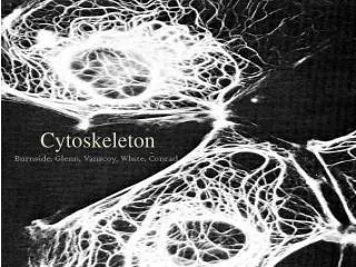
CYTOSOL AND CYTOSKELETON. CYTOSOL: fluid part of the cell cytoplasm Components: water ions enzymes inclusion bodies. CYTOSKELETON 3 classes of fibres: 1./ actin microfilaments (7 nm) 2./ intermediate filaments (10 nm) 3./ microtubules (25 nm)

E N D
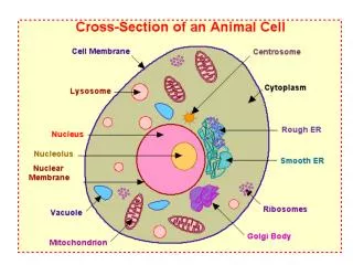
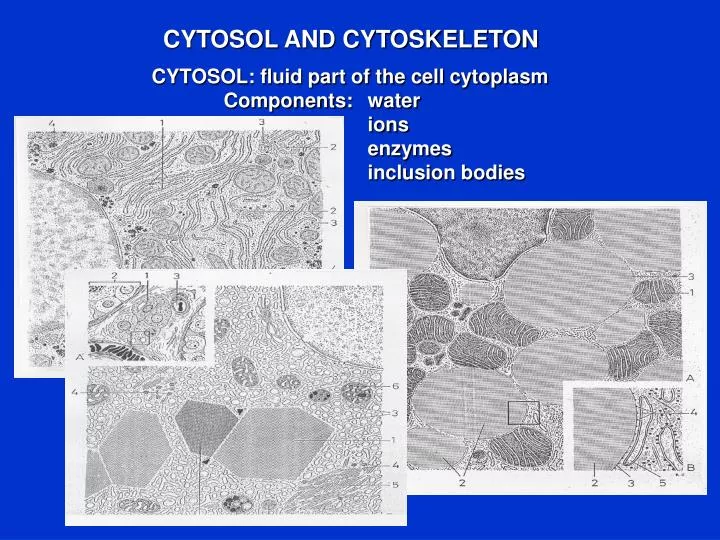
CYTOSOL AND CYTOSKELETON CYTOSOL: fluid part of the cell cytoplasm Components: water ions enzymes inclusion bodies
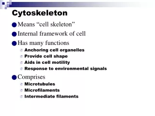
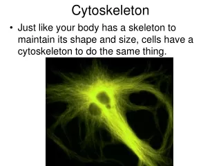
CYTOSKELETON 3 classes of fibres: 1./ actin microfilaments (7 nm) 2./ intermediate filaments (10 nm) 3./ microtubules (25 nm) Functions: maintainance of the cell shape take part in the cell motility serve as anchoring points
ACTIN MICROFILAMENTS I. They play a role in every type of cell motility. Types:a- actins (3 subtypes) non-muscle b-actin non-muscle g-actin g-actin in intestinal smooth muscle States of actin: globular (G) actin: ATP-G-actin (predominant) ADP-G-actin Fibrillar (F) actin: ATP-F-actin ADP-F-actin (predominant) Organization of actin cytoskeleton: bundles of filaments networks of filaments: planar 3-dimensional
Actin (green) and mitochondria (orange) in a fibroblast cell Molecular structure of a G-actin molecule Organization of G-actin into F-actin molecular complex
ACTIN MICROFILAMENTS II. Actin cross-linking proteins: short: fimbrin, a-actinin (bundles) long: filamin, spectrin, dystrophin (networks) Polymerization of actin filaments: 3 phases: lag phase: formation of a „nucleus” growth phase: elongation of the „nucleus” equilibrium between the amounts of G- and F-actin
MYOSIN • Motor protein, or mechanochemical enzyme. • Types and functions: • 1./ Myosin I.: cytoskeleton-membrane interactions • 2./ Myosin II.: muscle contraction and cytokinesis • 3./ Myosin V.: cytoskeleton-membrane interactions • Parts: head: actin- and ATP binding sites • neck: regulation of the activity of the head • tail:binding sites determining the formation of a dimer • Function: • contraction in muscle cells • stress fibres in non-muscle cells • stiffen cortical membranes • take part in cytokinesis Light chains Heavy chains Heads
MICROTUBULES I. Structure:a- and b-tubulins form heterodimers heterodimers form protofilaments protofilaments form a microtubule Cytoplasmic microtubules: stable, long-lived dynamic, short-lived Microtubule dynamics: they can oscillate between growing and shortening: dynamic instability
MICROTUBULES II. Microtubular cell organelles: cilia, flagella, basal body, centrioles
MTOC: microtubule organizing center: amorphous cytosol centrioles g-tubulin pericentrin centrioles g-tubulin pericentrin
INTERMEDIATE FILAMENTS: • They are smaller than microtubules, but larger than microfilaments • They are present in all eukaryotic cells (epithelial cells and neurons • contain the most) • They are key determinants of cellular structure • Functions: • To reinforce cells • To organize cells into tissues • Main properties: • Stability • Assembly of a-helical rods • Do not bind nucleotides
Types: 1./ Keratins: interconnect adjacent epithelial cells 2./ Neurofilaments: form the core of long axons 1./ 2./
3./ Glial filaments: present in astrocytes 4./ Vimentin: in fibroblasts and adipocytes 3./ 4./
Intermediate filaments can be useful in the diagnosis of cancer. Tumour cells loose their character and it is difficult to establish their origin. The identification of one of the intermediate filaments can identify their epidermal, neuronal, glial, or mesenchymal origin.