Download
1 / 36
360 likes | 685 Views
OSTEOMYELITIS-At-Shaheed-Suhrawardy-Medical-College-Hospital-Dhaka-Bangladesh

E N D
CME ON - OSTEOMYELITIS Basic of From - Department Of Orthopaedics & Traumatology. SheedSuhrawardy Medical College Hospital, Dhaka-1207, Bangladesh.
Presenting By- Dr. GolamMahamudSuhash, From Department Of Orthopedic & Traumatology, ShaheedSuhrawardy Medical College Hospital, Dhaka-1207. Bangladesh. OSTEOMYELITIS Prepared By- Dr. Md Nazrul Islam MBBS, M . sc. (B M E).
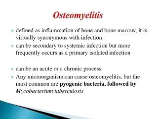
Definition of Osteomyelitis: Osteomyelitis may be defined as an acute or chronic inflammatory process of bone, bone marrow and its structure secondary to infection with micro organisms.
Staging Of Osteomyelitis: The Cierny-Mader staging system is used – It is determined by the status of the disease process regardless of its aetiology, regionality or chronicity. It takes into account the state of the bone, the patient's overall condition and factors affecting the development of osteomyelitis.
Anatomical Staging Of Osteomyelitis: 1: Medullaryosteomyelitis (infection confined to the bone surface) Stage. 2: Superficial osteomyelitis (contiguous type of infection)Stage. 3: Localisedosteomyelitis – (full-thickness cortical sequestration which can easily be removed surgically)Stage . 4: Diffuse osteomyelitis (loss of bone stability, even after surgical debridement).
Etiological agents: • In Newborns (younger than 4 mo) - S. aureus, Enterobacter species, andgroup A and B Streptococcus species • In Children (aged 4 mo to 4 y) - S. aureus, group A Streptococcus species, Haemophilus influenzae, and Enterobacter species • In Children, adolescents (aged 4 y to adult) - S. aureus (80%), group A Streptococcus species, H. influenzae, and Enterobacter species • In Adult - S. aureus and occasionally Enterobacter or Streptococcus species • In Sickle Cell Anemia Patients -Salmonella species • Fungus also causes osteomyelitis.
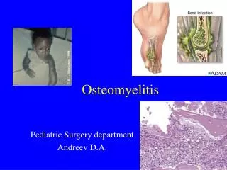
Hematogenous-common in children • Direct inoculation (usually traumatic, but also surgical) • Local invasion from a contiguous infection (usually decubitus ulcer or periodontal disease) Pathogenesis
Pathogenesis(cont….) • Hematogenousosteomyelitis highest in the first two decades of life. • < 5 years of age. • In adult-Haematogenous is less common but they suffered due to debility,disease(diabetes mellitus)drugs(immunosuppresion).
Pathogenesis(cont….) Discontinuous endothelium of distal metaphyseal vessels ----passage of bacteria through these gaps ---deposition • Trauma or emboli lead to occlusion of the slow-flowing sinusoidal vessels, further establishing a nidus for infection. • Metaphyseal capillaries--- lack phagocytic lining cells • Sinusoidal veins contain - inactive phagocytic cells. • Necrosis of cortical bone and marrow occurs. • Exudate under pressure is forced through the Haversian systems and Volkmann canals and into the cortex of the bone.
Pathogenesis(cont….) Pus spreads into the bone's blood vessels, impairing their flow, and areas of devitalized infected bone, known as sequestra, form the basis of a chronic infection.Often, the body will try to create new bone around the area of necrosis. The resulting new bone is often called an involucrum.Onhistologic examination, these areas of necrotic bone are the basis for distinguishing between acuteand chronicosteomyelitis.
Newborns- • Joint involvement is common • Nutrient metaphyseal capillaries perforate the epiphyseal growth plate, particularly in the hip, shoulder, and knee thin metaphyseal cortex.
Factors That Turn Acute Bone Infection to Chronic Osteomyelitis: Trauma (orthopaedic surgery or open fracture) Prosthetic orthopaedic device Diabetes Peripheral vascular disease Chronic joint disease Alcoholism Intravenous drug abuse Chronic steroid use Immunosuppression Tuberculosis HIV and AIDS Sickle cell disease Presence of catheter-related blood stream infection.
Diagnosis Signs and symptom • Temperature >102ºF • long-lasting pain, • Decreased range of motion in the case of joint involvement. • local warmth, tenderness, swelling
Diagnosis(cont….) • Aspiration of pus and send for culture • W.B.C. CRP and ESR • Blood for culture • Plain films, bone scintigram,ultrasound,CT Scan and MRI Even a biopsy all show positive results
Laboratory findings: • Elevations in the peripheral white blood cell count (WBC), • Erythrocyte sedimentation rate (ESR), and C-reactive protein (CRP) in children with hematogenousosteomyelitis are variable and nonspecific • Blood culture is positive in half of cases.
Lytic and sclerosis, indicating chronic infection. • Periostealnew bone formation, with compatible clinical findings Plain radiographs shows-
Withinthree to seven days- • interposed translucent fat planes within muscle are obliterated by edema fluid. • Periostealelevation or thickening may represent new bone formation, pus, or reactive edema from adjacent soft tissue infection
Timing • Age --- neonates, bone destruction is often apparent by the 7th to 10th day of illness • Type of bone ---Long tubular bones show signs two to three weeks earlier than membranous or irregular bones. • plain films of the pelvis -- not helpful
Timing Radiographic evidence of bone destruction finally becomes apparent by 10 to 21 days.
Magneticresonance imaging • Modality of choice • Accurate identification of subperiosteal or soft tissue collections of pus. no radiation. • Excellent anatomic detail and differentiation among soft tissue, bone marrow, and bone • signal from infected bone marrow can also be enhanced with intravenous gadolinium contrast – when plain films are normal
Scintigraphy • Sensitivity (84 to 100 percent) and specific (70 to 96 percent) • Usually readily available, • Relatively inexpensive, • Does not require sedation in young children. • May not perform as well in neonates • Accuracy in children in 90%
Computed tomography • CT preferred over plain films • Planning the surgical approach to debridement of sequestra • Evaluating the patient being treated for osteomyelitis
Differential diagnosis - • Cellulitis\ • Necrotizing myositis • Suppurative Arthritis • Rheumatism • Sickle cell crisis • Gaucher,s disease • Ewing,s sarcoma
ManagementAnd Treatment Of Osteomyelitis: General principles- • Early clinical suspicion, confirmation through imaging and microbiological tests and prompt treatment are the keys to a successful outcome. • Analgesia (and limb splinting if a long bone is involved) is an important part of symptom control. • Exact treatment varies according to the bones involved, the severity of the infection and the immune status of the patient. • Surgery may be needed to debride the bone and close any defects.
Management And Treatment Of Osteomyelitis (Cont.): • In acute osteomyelitis- The principle of treatment are- 1.General supportive treatment Analgesic for relieve pain I/V fluid(fever with shock,septicaemia) 2.Spintage of the affected part 3.Antibiotics(oral/intravenous)-It should be started immediately not waiting for culture of blood and pus
Management And Treatment Of Osteomyelitis: (con.) Antibiotics- • In older children who have probably s.aureus infection use I/V flucloxacillin and fusidic acid for 1 to 2 weeks then orally 3 to 6 wks. • In children under 4 years who have high incidence of haemophilus influenza use cephalosporins(cefuroxime or cefotaxime) either I/V or orally.Alternatively also use combination of amoxycillin and clavulanic acid 4.Drainage-if necessary
Complications of acute osteomyelitis • If treatment is delayed or the organism insensitive to the chosen antibiotics arise some complications- 1. Metastatic infection 2. Suppurativearthritis 3. Altered bone growth 4. Chronic osteomyelitis
Treatment(cont…) • In chronic osteomyelitis- Treatment options are- 1. Antibiotics- Chronic infection is seldom eradicated by antibiotics alone.Fusidicacid,clindamycin and cephalosporins are good examples. 2. Local treatment- If sinus present-need simply dressing Colostomy paste used to stopped excoriation of skin. In acute abscess-need urgent incision and drainage but it is temporary.
Management AndTreatmentOfOsteomyelitis (Cont.): 3. Operation-procedures are: • 1.Saucharization and curettage • 2.Gentamycin impregnated beads • 3.Papineau technique and muscle flap transfer. • 4.Ilizarov method of bone transporting.
MONITORING RESPONSE TO THERAPY • A reduction in fever and pain, as well as increased comfort with movement are expected within seven days, and may be seen in toddlers in as little as three to five days. • Persistent elevation or prolonged decline in ESR or CRP. The ESR should have substantially decreased (by 20 percent or more) by the end of the first week of therapy, with a 50 percent decline in CRP. • continue to decline during the second week of treatment. CRP returns to normal in 10 days or less in most patients.
OUTPATIENT THERAPY- • Treatment with intravenous antibiotics for periods of seven days or less, followed by oral therapy, appears to be as successful as longer initial parenteral courses • Followup one- to two-week intervals --monitored clinical improvement, -- complications related to high-dose antibiotic therapy, such as cytopenia, antibiotic associated diarrhea, and pseudomembranousenterocolitis. • ESR, complete blood count, and biochemical profile, including liver function tests at each visit. drug levels for those children receiving oral therapy • A radiograph should be repeated at the end of therapy.
Most CommonComplications Of Acute Osteomyelitis: • Bone abscess • Septic Arthritis • Bacteraemia • Fracture • Growth arrest • Loosening of the prosthetic implant • Overlying soft-tissue cellulitis • Chronic infection
Prognosis Of Osteomyelitis: • This is variable depending on the number of risk factors and the patient's general condition (see Staging above). • Outcome is best if treatment is started 3-5 days after onset of the infection. • Timely diagnosis and intervention in an otherwise well patient should lead to full recovery, although follow-up over several months will be required to monitor for relapse.
General Guide Lines For Evaluation & Management of Osteomyelitis:
InceptaPharmaceuticals Ltd.Dhaka, Bangladesh. Prof. Dr. ShamimulHaq Associate Prof. Dr. P C. Debenath Associate Prof. Dr. ZiaulHaq Associate Prof. Dr. HafizurRahman Assistant Prof. Dr. KaziShamimuzzaman Special Thanks to - &