Download

1 / 45
460 likes | 664 Views
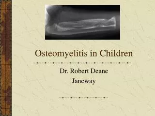
Osteomyelitis. Epidemiology. Osteomyelitis and suppurative arthritis are most common in young children 1/3 cases < 2 y/o 1/2 cases < 5 y/o boys : girls, usually 2:1 1/3 cases have minor, closed trauma. Etiology. A microbial etiology is confirmed in about 3/4 of cases of osteomyelitis

E N D
Epidemiology. • Osteomyelitis and suppurative arthritis are most common in young children • 1/3 cases < 2 y/o • 1/2 cases < 5 y/o • boys : girls, usually 2:1 • 1/3 cases have minor, closed trauma
Etiology. • A microbial etiology is confirmed in about 3/4 of cases of osteomyelitis • Staphylococcus aureus • the most common infecting organism in all age groups • Group B streptococcus and gram-negative enteric bacilli • prominent pathogens in neonates • group A streptococcus • less than 10% of all cases • After 6 yr of age, most are S. aureus
Etiology. • Exclusive. • puncture wounds of the foot: Pseudomonas aeruginosa • sickle cell anemia: Salmonella and S. aureus • other consideration: • penetrating injuries: atypical mycobacteria • Fungal infections usually occur as part of multisystem disseminated disease • Candida osteomyelitis sometimes occursin neonates with indwelling vascular catheters • Primary viral infections of bones--> rare; • but complain of arthralgia or arthritis may be due to immune responce
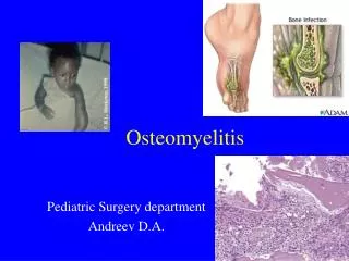
Pathogenesis • In newborns and young infants: • transphyseal blood vessels connect the metaphysis and epiphysis • common for pus from the metaphysis joint space • latter part of the first year of life, the physis forms • obliterating the transphyseal blood vessels • later childhood the periosteum becomes more adherent, • favoring pus to decompress through the periosteum • growth plate closes in late adolescence • begins in the diaphysis, and can spread to the entire intramedullary canal
Pathogenesis. • In the metaphysis, nutrient arteries branch into nonanastomosing capillaries under the physis, which make a sharp loop before entering venous sinusoids draining into the marrow • bacterial focus established --> phagocytes --> inflammatory exudates --> metaphyseal space--Halverson system and Volkmann canals) --> subperiosteal space --> impairing blood supply to the cortex and metaphysis
Clinical Manifestations. • signs and symptoms highly dependent on the age, • earliest signs and symptoms are often subtle • latent period of 10 to 12 days between the time of onset of clinical symptoms and the development of definite radiographic changes in bone. • Neonates: • pseudoparalysis • Half of do not have fever , may not appear ill
Clinical Manifestations. • Older infants and children • fever, pain, and localizing signs such as edema, erythema, and warmth • limp or refusal to walk • Local swelling and redness with osteomyelitis may mean that the infection has spread out of the metaphysis into the subperiosteal space, representing a secondary soft tissue inflammatory response
Clinical Manifestations. • upper extremities account for one fourth of all cases. Flat bones are less commonly • Several bones or joints are infected in fewer than 10% of cases • exceptions are gonococcal infections and osteomyelitis in neonates • two or more bones are involved in almost half of the cases
Diagnosis. • Take samples: • Aspiration of the infected site for Gram stain and culture • steel needle is needed to penetrate the cortex into the metaphysis. • If pus is encountered in the subperiosteal space, there is no need to go farther. • If gonococcus is suspected, • cervical, anal, and throat cultures should also be obtained • blood culture should be performed
Diagnosis. • Other Lab. Exam • no specific laboratory tests for osteomyelitis • CBC/DC, ESR, CRP--> very sensitive but not specific • normal test results cannot r/o osteomyelitis • can monitor dz progress
Diagnosis. • RADIOGRAPHIC EVALUATION. • Plain Radiographs. • Within 72 hours of onset of symptoms of osteomyelitis • soft tissue technique, displacement or obliteration of the normal fat planes adjacent to deep muscle • compared to the opposite extremity, • can show displacement of the deep muscle
Diagnosis. • Examination obtained 10 days after the onset • B: Repeat examination 1 week
Diagnosis. • Plain Radiographs. • minimal amount of periosteal new-bone formation laid down parallel to the outer margin of the cortex • The actual disease process is usually much more extensive than showed by radiograph
Diagnosis. • Examination 7 days after the onset • 3 months later shows extensive new-bone formation
Diagnosis. • Ultrasonography. • detecting joint effusion and fluid collection in the soft tissue and subperiosteal regions • may guide localization for aspiration or drainage.
Diagnosis. • Computed Tomography and Magnetic Resonance Imaging. • CT is ideal for detecting gas in soft tissues • Increased attenuation occurs within the bone marrow early in the disease due to edema and pus
Diagnosis. • MRI is the best radiologic imaging technique for the identification of abscesses and for differentiation between bone and soft tissue infection • acute osteomyelitis, purulent debris and edema appear dark with decreased signal intensity on T1 weighted images opposite is seen in T2 weighted images • for possible surgical intervention • MRI is particularly useful in the evaluation of vertebral osteomyelitis and diskitis clear delineation
Diagnosis. • MRI • MRI does not reliably distinguish osteomyelitis from noninfectious bone marrow edema • not sensitive to changes within the cortex
Diagnosis. • Radionuclide Studies. • be valuable especially if multiple foci are suspected • Technetium-99 methylene diphosphonate (99m Tc), which accumulates in areas of increased bone turnover • 99mTc 3 phase perfusion, blood pool, and delayed images • Any areas of increased blood flow or inflammation can cause increased uptake of 99m Tc in the • first phase (5–10?min) and • second phase (2–4?hr), • but osteomyelitis causes increased uptake of 99m Tc in the third phase (24?hr)
Diagnosis. • Radionuclide Studies. • sensitivity (84–100%) and specificity (70–96%) • can detect osteomyelitis within 24–48?hr after onset of symptoms • Gallium-67 uptake, Indium-111 leukocytes is more specific for infection • sensitivity in neonates is much lower owing to poor bone mineralization
Treatment. • Optimal treatment of skeletal infections requires collaborative efforts of pediatricians, orthopedic surgeons, radiologists, and physiatrists • one should not wait for the development of radiographic evidence of disease before treatment • empirical antibiotic • antistaphylococcal penicillin, such as nafcillin or oxacillin, and a broad-spectrum cephalosporin, such as cefotaxime • aminoglycoside may be take place of cephalosporin • but reduced antibacterial activity in sites with decreased oxygen tension and low pH
Treatment • In infants and children younger than 5 yr of age • Cefuroxime • After 5 yr of age and in the absence of special circumstances • nafcillin or cefazolin • With sickle cell disease , gram-negative enteric bacteria are common • broad-spectrum cephalosporin such as cefotaxime or ceftriaxone
Treatment • When the pathogen is identified, appropriate adjustments in antibiotics are made • If a pathogen is not identified and a patient's condition is not improving • re-aspiration or biopsy • Recheck diagnosis
Treatment • Duration of antibiotic (1) the patient shows prompt resolution of signs and symptoms (within 5–7 days) and (2) the ESR has normalized; a total of 4–6 wk of therapy may be required • S. aureus or gram-negative bacillary infections, the minimum duration of antibiotics is 21 days • group A streptococcus, S. pneumoniae, or H. influenzae type b, antibiotics minimum of 10–14 days • Pseudomonas postoperative need total of 7 d treatment • Immunocompromised, mycobacterial or fungal infection patients generally require prolonged courses
Treatment • Oral route • Changing antibiotics from the intravenous route to oral administration when a patient's condition has stabilized, generally after 1 wk • serum bactericidal titers, or Schlichter titers, 45–60?min after a dose of suspension or 60–90?min after a capsule or tablet. • A serum bactericidal titer of 1:8 or more is considered desirable • ß-lactam drugs for staphylococcal or streptococcal infection, a dosage 2~3 times that used for other infections is prescribed
Treatment • SURGICAL THERAPY. • When frank pus is obtained from subperiosteal or metaphyseal aspiration a surgical drainage procedure is usually indicated. • penetrating injury and when a retained foreign body • Infection of the hip is considered a surgical emergency • chronic osteomyelitis consists of surgical removal of sinus tracts and sequestrum Antibiotic therapy is continued for several months until clinical and radiographic
Prognosis. • Failure to improve or worsening by 72?hr requires review of the appropriateness of the antibiotic therapy • CRP typically normalizes within 7 days after start of treatment; ESR typically rises for 5–7 days, then falls slowly; dropping sharply after 10–14 d • Recurrence of disease and development of chronic infection after treatment < 10% of patients • initiation of medical and surgical therapy < 1 wk of onset of symptoms better prognosis
SUBACUTE AND CHRONIC OSTEOMYELITIS • Bone Abscess • lucency area surrounded by an irregular zone of dense sclerosis. • The overlying cortex is usually thickened by periosteal new-bone formation
SUBACUTE AND CHRONIC OSTEOMYELITIS • Involucrum and Sequestrum Formation • Sequestra become avascular by losing their • periosteal supply: periosteum is stripped, elevated from cortex • intramedullary supply: infection causes vascular thrombosis • areas of dense bone surrounded by zones of lucency
SUBACUTE AND CHRONIC OSTEOMYELITIS • involucrum is a shell of bone formed by the periosteum that surrounds and encloaks a sequestrum • Involucrum and sequestrum formation in osteomyelitis more common in children than in adults • MRI: • focus of active infection is similar to that of a bone abscess, low on T1, higher than marrow on T2, surrounded by a low-intensity rim on both
DIFFERENTIAL DIAGNOSIS. • trauma • Neuroblastoma • Primary bone tumors • Cellulitis • DVT
Cellulitis • skin infection, soft-tissue swelling is superficial and does not involve the deeper tissues adjacent to the bone.
Neuroblastoma • arises from sympathetic nervous tissue, often within the adrenal gland. • A palpable mass in the abdomen may be the first evidence • metastatic neuroblastoma is the most common malignant-appearing tumor before the age of 1 year in bone tumor • Calcification often is visible within the primary tumor
Neuroblastoma • X-ray • cranial sutures are spread • In the skull: Thin, whisker-like calcifications frequently extend out and inward • long tubular bones: • moth-eaten, and the cortex often eroded • Periosteal new-bone formation may parallel the cortex or form thin spiculations at right angles to the cortex • Ewing's tumor, neuroblastoma, and leukemia, may have a similar radiographic appearance, particularly in the long bones.
Ewing's tumor • malignant tumor arising in the red bone marrow and closely related histologically to reticulum cell sarcoma • Mostly: 5 ~ 25 y/o ; rarely after 30 y/o; male> female • long bones, extremities, femur, metaphysis • Exception: > 20 y/o, flat bones > long bones • Metastasize easy, multiple lesions may be present • symptoms : pain, swelling; often fever and leukocytosis, like osteomyelitis
Ewing's tumor • (L) Laminated, onionskin, periosteal new-bone formation • (R) permeative destruction and a well-defined, laminated Codman's triangle
Ewing's tumor • X-ray • permeative, poorly marginated, destructive lesion that perforates the cortex • overlaid by a laminated, , onionskin periosteal reaction • Others may find: Codman's triangles, Fine spicules, • In the long bones, differentiation of Ewing's tumor and osteomyelitis may be difficult