Download

1 / 27
490 likes | 1.45k Views
EXAMINATION OF THE EYE. DR.M.P.LAL. PROFESSOR AND HEAD OF DEPT OF SURGERY. HISTORY. PRESENT HISTORY. Name ,Age,Sex ,Occupation. Dimness of vision . Pain in eyes Redness , congestion or inflammation Secretion Disturbances of the eyeball Headache. Past history.

E N D
EXAMINATION OF THE EYE DR.M.P.LAL. PROFESSOR AND HEAD OF DEPT OF SURGERY
PRESENT HISTORY • Name ,Age,Sex ,Occupation. • Dimness of vision . • Pain in eyes • Redness , congestion or inflammation • Secretion • Disturbances of the eyeball • Headache
Past history • Previous diseases . • Treatment . • Operation. • History of use of glasses .
Personal history • Habits • Blood pressure • Diabetes mellitus • Renal, haematologic and cardiac diseases • Foci of infection in teeth,tonsils,ears and sinuses.
Family history • Diabetes mellitus • Hypertension • Myopia • Glaucoma • Congenital cataract
Examination of anterior segment of eye • INSPECTION • PALPATION • INTRAOCULAR TENSION • BINOCULAR LOUPE AND SLIT – LAMP EXAMINATION • GONIOSCOPE EXAMINATION • TRANSIILLUMINATION.
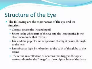
1.Inspection in anterior segment GENERAL INSPECTION OF - HEAD /FACE /ORBITS EYEBALLS / EYELIDS LACRIMAL SAC/ CORNEA CONJUNCTIVA / SCLERA ANTERIOR CHAMBER / IRIS PUPIL/ LENS
3.INTRAOCULAR TENSION NORMAL INTRAOCULAR PRESSURE- 10-20 mm hg (schiotz) SUSPICIOUS CASES = 20-25 mmhg (schiotz) GLAUCOMA = above 25 mmhg(schiotz) METHODS • DIGITAL TENSION • SCHIOTZ TONOMETER • APPLANATION TONOMETER –
4.BINOCULAR LOUPE AND SLIT – LAMP EXAMINATION Examination of eye is done in focal or oblique illumination under magnification. BINOCULAR LOUPE – A stereoscopic effect is obtained & depth of opacities can be assessed. MAGNIFICATION – 3-4 times SLIT – LAMP EXAMINATION – Essential when minute examination is essential. MAGNIFICATION – 16 – 25 times.
5.GONIOSCOPIC EXAMINATION ABNORMAL ANGLE STRUCTURES ARE IDENTIFIED. TYPES- • Direct with goniolenses • Indirect with gonioprisms NORMAL ANGLE STRUCTURES Schwalbe’s line , Trabecular meshwork , Scleral Spur & Ciliary band.
6.transillumination • TRANS-SCLERAL – when an intense beam of light is thrown through sclera , pupil appears red in colour. The pupil remains black if there is solid mass. • TRANS-PUPILLARY- when an intense beam of light is passed obliquely through dilated pupil , pupil becomes illuminated uniformly in normal cases.
EXAMINATION OF THE POSTERIOR SEGMENT OF EYE 1.EXAMINATION OF RETINAL FUNCTIONS • SUBJECTIVE EXAMINATION • OBJECTIVE EXAMINATION 2.EXAMINATION OF THE FUNDUS OCULI
I.VISUAL ACUITY • SNELLEN’S TEST TYPE 1.RECORDING FOR DISTANCE- Normal visual acuity = 6/6 OTHER TEST TYPE- a.LANDOLT’S CHART b.E CHART c.SIMPLE PICTURE CHART. 2.RECORDING FOR NEAR-normal vision is recorded as N/6
II. Field of vision NORMAL FIELD OF VISION- • UPWARDS – 60º/INWARDS - 60º • DOWNWARDS - 70º • OUTWARDS – MORE THAN 90º TECHNIQUES EMPLOYED TO EVALUATE BOTH CENTRAL & PERIPHERAL VISUAL FIELDS IS PERIMETRY a.KINETIC PERIMETRY b.STATIC PERIMETRY
FIELD OF VISION PERIPHERAL FIELD • CONFRONTATION METHOD • THE PERIMETER (Lister’s, Goldmann’s) CENTRAL FIELD • BJERRUM’S SCREEN • AUTOMATED PERIMETERS
III. Colour vision OBJECTIVES- • to find out the exact nature of testing colour vision. • Whether the patient is likely to be a source of danger to the society.eg .driver,pilot,sailor etc
Examination of fundus oculi NEXT TO MEDIA- optic disc macula lutea retinal vessels general fundus
Examination of fundus by focal illumination FOUR TYPES OF LENSES ARE AVAILABLE FOR BIOMICROSCOPIC EXAMINATINATION OF THE VITREOUS AND FUNDUS: • HRUBY’S LENS • POSTERIOR FUNDUS CONTACT LENS • GOLD MANN THREE MIRROR CONTACT LENS • INDIRECT SLIT LAMP BIOMICROSCOPY USING +78D,+90D
ANCILLARY INVESTIGATIONS STRUCTURES THAT ARE NOT EASILY VISIBLE BY DIRECT OBSERVATION CAN BE STUDIED BY; • FLUORESCEIN ANGIOGRAPHY • INDOCYANINE ANGIOGRAPHY • ULTRASONOGRAPHY • OPTICAL COHERENCE TOMOGRAPHY(OCT) • SCANNING LASER OPHTHALMOSCOPY-COMPUTERIZED AXIAL TOMOGRAPHY(CAT) • RADIOLOGICAL INVESTIGATIONS – (MRI) • OPHTHALMO-DYNAMOMETRY