Download
1 / 46
640 likes | 1.96k Views
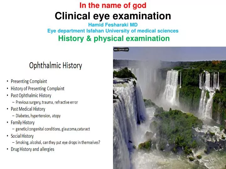
In the name of god Clinical eye examination Hamid Fesharaki MD Eye department Isfahan University of medical sciences History & physical examination. Clinical eye examination History & physical examination. Ophthalmic symptoms : pain, redness, itching, burning , F B sensation, Visual loss.

E N D
In the name of god Clinical eye examinationHamid Fesharaki MDEye department Isfahan University of medical sciences History & physical examination
Ophthalmic symptoms : pain, redness, itching, burning , F B sensation, Visual loss Clinical eye examinationHistory & physical examination Snellen Chart Hand Held Acuity Card
Physical examinationvisual acuity: fixation & follow, snellen chart
Clinical examination Visual acuity: central, peripheral visual acuity is hard to check due to its subjective nature: depends on the response of the patient (intelligence , previous experience, alertness)
Measurment of visual acuitymonocular vs binocular, wit or without correctionfar and near
Accurate clinical eye examinationreduces the para clinical expensive testingPoor ophthalmoscopy may call for ocular sonography, OCT, FA…Define the best corrected visual acuity firstRefraction is the beginning step of clinical examination clinical judgment without refraction can be miss guiding RAPD (Retrobulbar neuritis)Judgment by observation alone may be misleading A relatively pale optic disc Reduced light reflex of fovea Optometrist referral for retinal problem
Refractive error: retinoscopy, subjective refraction including the pin hole , autorefraction.
subjective refraction To find the best corrected visual acuity
Observation of the fundus structures is very important for clinical diagnosis.
Visual loss: 1. Refractive error: retinoscopy, subjective refraction including the pin hole , autorefraction. (Irregular astigmatism) 2. Opacity of media: ophthalmoscopy, retinoscopy, biomicroscopy. (red reflex) 3. Retina & Optic nerve : ophthalmoscopic observation, RAPD, visual field, ERG, EOG, VEP, angiography, OCT, ultrasonography. (Amblyopia) Amblyopia: history & phsical:Anisometropia, Isoametropia, Strabismic, (Monofixation synd) 4 prism base out test Malingering: age, gain, tricksLegal writing Beyond the optic nerve: RAPD,VEP,Visual field, brain imaging Deprivation
. Opacity of media: ophthalmoscopy, retinoscopy, biomicroscopy. ultrasonography (red reflex)
Evaluation of retina & optic nerveVisual field: Confrontation, tangent screen
Evaluation of retina & optic nerve • Observation: Compare between the two eyes, and compare with the population. • Correlate between BCVA, clarity of visual pathway and fundascopic findings • Relative afferent pupillary defect • Function tests: visual field,VEP, ERG, EOG..
Retina & Optic nerve :Angiography, visual field, OCT, GDX, ERG, EOG, VEP, ultrasonography,
Beyond the optic nerve: Visual field brain imaging: MRI, CTscan
Observation External StructuresPupil, iris and eyelids & lashes should appear symmetricSclera should be whiteConjunctiva clear