Download

1 / 43
620 likes | 1.22k Views
Microbial Growth. Chapter 6. Growth Requirements. Physical Temperature Optimum growth temperature- temperature at which the organism grows its best Psychrophiles - cold loving- range from –10 º C to < 20 º C; optimum 15 º C

E N D
Microbial Growth Chapter 6 Dr. G. López-de-Victoria
Growth Requirements • Physical • Temperature • Optimum growth temperature- temperature at which the organism grows its best • Psychrophiles- cold loving- range from –10 ºC to < 20 ºC; optimum 15 ºC • Mesophiles- moderate-temperature-loving- optimum 25-40 ºC; most common • Thermophiles- heat-loving- optimum 50-60 ºC • Psychotrophs- range from 0 ºC to < 40 ºC; responsible for food spoilage GLV
Growth Requirements (cont.) • pH- most bacteria grow best between pH 6.5 and 7.5; fungi prefer more acidic environments, optimum pH, 5-6 • Acidophiles- acid loving; can live as low as pH 1, e.g. coal mines • Osmotic pressure • Halophiles- salt-loving- some can grow in as much as 30% NaCl GLV
Fig. 6.3 The effect of the amount of food on its cooling rate… GLV
Chemical • Water • Carbon sources • Heterotrophs-organics • Autotrophs- CO2 • Sources of • Nitrogen- amino acids, nucleic acids • Sulfur- amino acids, vitamins • Phosphorous- ATP GLV
Chemical (cont.) • Trace elements- cofactors; iron, copper, zinc • Oxygen- Obligate aerobes • Anaerobes • Facultative anaerobes- use oxygen but can grow without it by fermentation or anaerobic respiration; have catalase and SOD, e.g. E. coli • Obligate anaerobes- lack enzymes to neutralize toxic forms of oxygen • Aerotolerant anaerobes- tolerates oxygen; SOD • Microaerophilic aerobes- low oxygen concentration • Enzymes- • Organic growth factors- e.g. vitamins, purines GLV
Enzymes • Superoxide dismutase- SOD- convert the superoxide free radical into molecular oxygen and hydrogen peroxide • Catalase- converts hydrogen peroxide into water and oxygen • Peroxidase- breaks down hydrogen peroxide into water GLV
Culture Media • Types • Agar- solid or semisolid • Plates, test tubes (deep or slanted) • Broth- liquid- NO agar • Chemically defined media- media whose exact composition is known • Useful for “fastidious” organisms • Complex media- nutrients derived from a variety of extracts; can vary slightly from one batch to the next • Nutrient broth- NB • Nutrient agar- NA GLV
Anaerobic Growth Media • Reducing media- contains ingredients such as sodium thioglycolate to help deplete the oxygen from the culture medium • Anaerobe jar- contains chemicals in a packet that generate hydrogen and carbon dioxide; when mixed with water the hydrogen then reacts with the oxygen present in the air and forms water, thus removing oxygen from the jar • Anaerobe chamber- equipped with air locks and gases; used when handling a lot of cultures GLV
Special Culture Techniques • Capnophiles- microorganisms that grow better at high CO2 concentrations • Carbon dioxide incubators • Candle jar GLV
Selective Media- used to suppress the growth of unwanted bacteria and encourage the growth of the desired organism • Bismuth sulfite agar • Inhibits Gram+ and most G- bacteria • Used to isolate Salmonella typhi • Sabouraud glucose agar • has a pH of 5.6 which inhibits most bacteria • used to isolate fungi • Brilliant Green Agar • inhibits G+ • Used to isolate Salmonella spp. GLV
Differential Media • Medium that makes it easier to distinguish colonies of the desired organism • Blood agar • used to identify organisms that lyse RBCs • E.g. Streptococcus pyogenes GLV
Fig. 6.8 Blood agar, a differential medium containing RBCs. GLV
Selective and Differential Media • Mannitol salt agar (MSA) • contains 7.5% NaCl- inhibitory • contains mannitol- carbon source • contains pH indicator that changes color if acid is produced from the fermentation of mannitol • organisms that can tolerate high concentrations of salt (selective component) and ferment mannitol (differential component) are likely to be Staphylococcus aureus GLV
MacConkey agar • Contains bile salts and crystal violet which inhibits G+ bacteria (selective) • Contains lactose (differential) • lactose fermenters appear as pink colonies; e.g. E. coli • non-lactose fermenters appear as colorless colonies GLV
Enrichment Media • Provides nutrients and environmental conditions that favor the growth of a particular microorganism and not others; encourages the growth of all bacteria present in a sample GLV
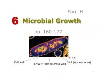
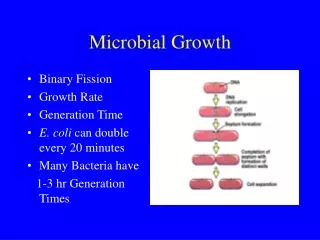
Cultures • Pure cultures • Colony- arises from a single cell or groups of cells • Streak Plate method- used to isolate colonies • Preservation • Deep-freezing- liquid suspension frozen –50 ºC to –70 ºC; lasts for years • Lyophilization- freeze-drying- suspension is quickly frozen and then sublimated- powder like product lasts for years and the suspension can be hydrated to “revive” the microorganism • Growth • Bacterial division- binary fission • Generation time- the time required for a cell to divide • Logarithmic growth GLV
Growth Curve • Lag phase- period of little or no division • Log phase • logarithmic increase in the number of bacteria; exponential • Stationary phase • The number of bacteria being produced equals the number of bacteria dying • Death or decline phase • Logarithmic decrease in the number of bacteria GLV
Measurement of Growth • Plate counts- measures viable cells; CFU- colony forming units • Serial dilutions- uses diluted samples • Pour plates and spread plates- • Filtration- used for samples with low microbial numbers; filter concentrates the microorganism • Most Probable Number (MPN) • Direct microscopic counts- direct enumeration of the cells GLV
Estimating Bacterial Numbers by Indirect Methods • Turbidity • Metabolic activity • Dry weight • Urine culture colony estimates’ • 10,000 or below- contamination • 10,000-100,000- not conclusive • > 100,000 - infection GLV