Download
1 / 6
80 likes | 90 Views
Based on Brownian motion, diffusion statement of proton hydro inside body is one of the most important variables affected on the diagnosis, treatment planning, and disease response to treatment. There are some different kinds of diffusion-weighted magnetic resonance imaging types such as diffusion-weighted imaging, diffusion tensor imaging, diffusion kurtosis imaging, intravoxel incoherent motion imaging, zoom diffusion imaging, and diffusion spectrum imaging. In this short communication, we aimed to introduce clinical applications of these diffusion-weighted magnetic resonance imaging types.

E N D
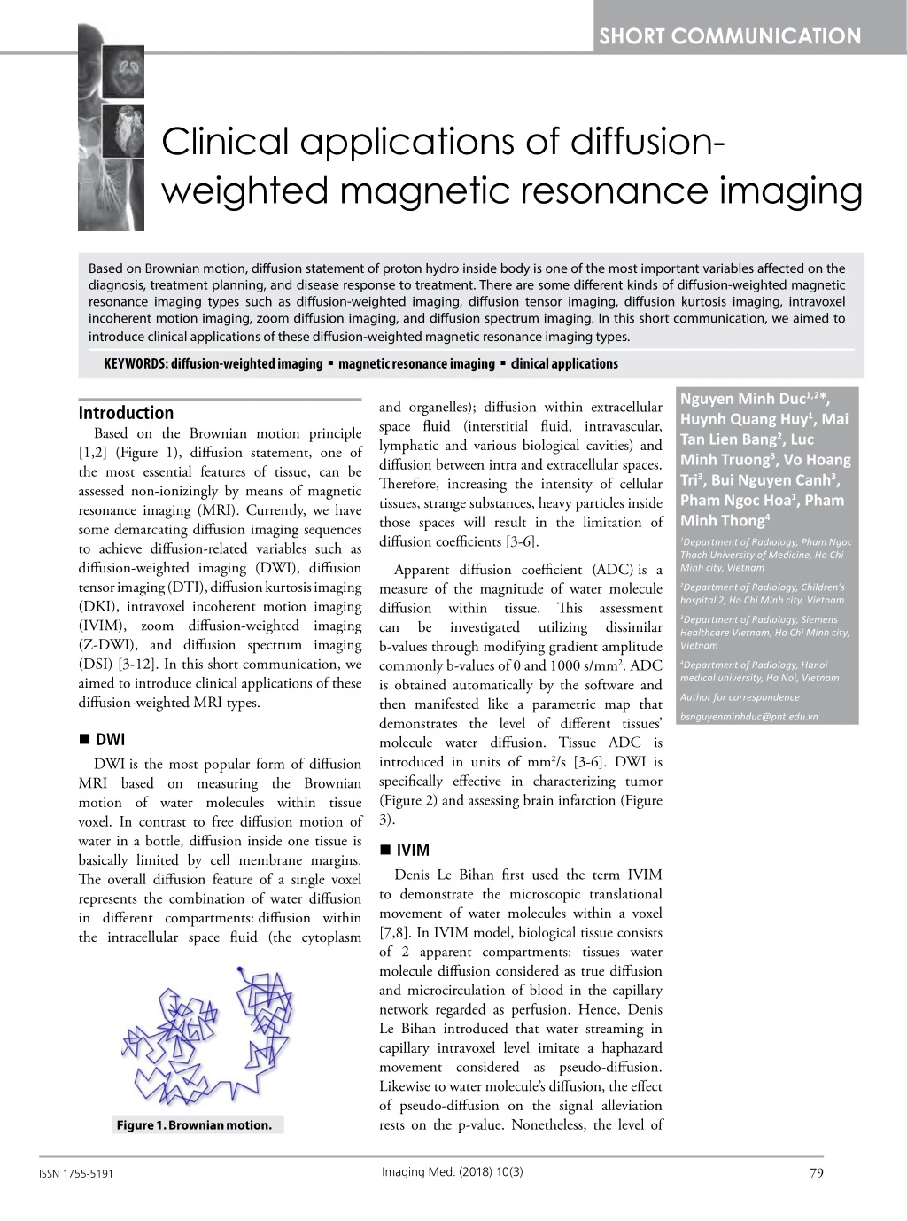
SHORT COMMUNICATION Clinical applications of diffusion- weighted magnetic resonance imaging Based on Brownian motion, diffusion statement of proton hydro inside body is one of the most important variables affected on the diagnosis, treatment planning, and disease response to treatment. There are some different kinds of diffusion-weighted magnetic resonance imaging types such as diffusion-weighted imaging, diffusion tensor imaging, diffusion kurtosis imaging, intravoxel incoherent motion imaging, zoom diffusion imaging, and diffusion spectrum imaging. In this short communication, we aimed to introduce clinical applications of these diffusion-weighted magnetic resonance imaging types. KEYWORDS: diffusion-weighted imagingmagnetic resonance imagingclinical applications Nguyen Minh Duc1,2*, Huynh Quang Huy1, Mai Tan Lien Bang2, Luc Minh Truong3, Vo Hoang Tri3, Bui Nguyen Canh3, Pham Ngoc Hoa1, Pham Minh Thong4 and organelles); diffusion within extracellular space fluid (interstitial fluid, intravascular, lymphatic and various biological cavities) and diffusion between intra and extracellular spaces. Therefore, increasing the intensity of cellular tissues, strange substances, heavy particles inside those spaces will result in the limitation of diffusion coefficients [3-6]. Introduction Based on the Brownian motion principle [1,2] (Figure 1), diffusion statement, one of the most essential features of tissue, can be assessed non-ionizingly by means of magnetic resonance imaging (MRI). Currently, we have some demarcating diffusion imaging sequences to achieve diffusion-related variables such as diffusion-weighted imaging (DWI), diffusion tensor imaging (DTI), diffusion kurtosis imaging (DKI), intravoxel incoherent motion imaging (IVIM), zoom diffusion-weighted imaging (Z-DWI), and diffusion spectrum imaging (DSI) [3-12]. In this short communication, we aimed to introduce clinical applications of these diffusion-weighted MRI types. 1Department of Radiology, Pham Ngoc Thach University of Medicine, Ho Chi Minh city, Vietnam Apparent diffusion coefficient (ADC) is a measure of the magnitude of water molecule diffusion within tissue. This assessment can be investigated b-values through modifying gradient amplitude commonly b-values of 0 and 1000 s/mm2. ADC is obtained automatically by the software and then manifested like a parametric map that demonstrates the level of different tissues’ molecule water diffusion. Tissue ADC is introduced in units of mm2/s [3-6]. DWI is specifically effective in characterizing tumor (Figure 2) and assessing brain infarction (Figure 3). 2Department of Radiology, Children’s hospital 2, Ho Chi Minh city, Vietnam 3Department of Radiology, Siemens Healthcare Vietnam, Ho Chi Minh city, Vietnam utilizing dissimilar 4Department of Radiology, Hanoi medical university, Ha Noi, Vietnam Author for correspondence bsnguyenminhduc@pnt.edu.vn DWI DWI is the most popular form of diffusion MRI based on measuring the Brownian motion of water molecules within tissue voxel. In contrast to free diffusion motion of water in a bottle, diffusion inside one tissue is basically limited by cell membrane margins. The overall diffusion feature of a single voxel represents the combination of water diffusion in different compartments: diffusion within the intracellular space fluid (the cytoplasm IVIM Denis Le Bihan first used the term IVIM to demonstrate the microscopic translational movement of water molecules within a voxel [7,8]. In IVIM model, biological tissue consists of 2 apparent compartments: tissues water molecule diffusion considered as true diffusion and microcirculation of blood in the capillary network regarded as perfusion. Hence, Denis Le Bihan introduced that water streaming in capillary intravoxel level imitate a haphazard movement considered as pseudo-diffusion. Likewise to water molecule’s diffusion, the effect of pseudo-diffusion on the signal alleviation rests on the p-value. Nonetheless, the level of Figure 1. Brownian motion. 79 Imaging Med.(2018) 10(3) ISSN 1755-5191
SHORT COMMUNICATION Duc, Huy, Bang, et al. Figure 2. Image findings show invasive ductal carcinoma in the left breast (white arrow) with restricted diffusion on DWI (left section) and ADC map (right section) in comparison with reference. Figure 3. Image findings show an infarction lesion of pons (white arrow) with restricted diffusion on DWI (left section) and color ADC map (right section) in comparison with reference. Figure 4. IVIM images show hepatocellular carcinoma (white arrow): (A) D-slow image prior to TACE; (B) D-fast image prior to TACE; (C) f-image prior to TACE; (D) D-slow image after TACE; (E) D-fast image after TACE; (F) f-image after TACE. Figure courtesy of Lin et al. [12]. signal alleviation caused by pseudo-diffusion is generally an order of magnitude larger than tissue’s molecular diffusion, therefore its respective addition to the DWI signal becomes important only at a very low b-value generally under b-value of 200 s/mm2, conceding diffusion and perfusion effects to be demarcated. In practice, IVIM was utilized to attain 4 parameters: F (perfusion fraction); D* (pseudo- diffusion); D (real-diffusion), and ADC (Figure 4). IVIM is very useful in assessing tumor features and tumor responses to therapy [7-12]. Z-DWI Conventional sophisticated structure generally produces not enough spatial resolution. In addition, conventional DWI is very sensitive to body movement and paramagnetic structure led to the geometrical distortion. So as to overcome this problematic condition, Z-DWI is a novel form of conventional DWI combined with high resolution even small field of view which can help clinician to investigate the lesions of sophisticated structure such as hippocampus, DWI when assessing 80 Imaging Med.(2018) 10(3)
SHORT COMMUNICATION Clinical applications of diffusion-weighted magnetic resonance imaging spinal cord or early stage of astrocytoma occurred in a small gyrus (Figure 5). Z-DWI is clinically indicated for epilepsy, characterization of hippocampus lesion, spinal cord lesion and assessment of brain tumor [13-15]. parameters: ADC and fractional anisotropy (FA) also known as diffusion anisotropy (Figure 7). In practice, the application of DTI is for brain such as tract-specific localization of white matter lesions, localization of tumors in relation to the white matter tracts, localization of the main white matter tracts for neurosurgical planning and assessment of the white matter maturation; nevertheless, today DTI can be applied to some other organs such as kidney, uterus, muscle and heart [16-19]. DTI DTI is an expansion of DWI that permits data contouring depended on tissue tract specifically white-matter The axon architecture in parallel bundles with outside myelin armor eases the water molecule’s diffusion along the same main direction (Figure 6) [16-19]. Fiber-tracking, also known as tractography, bases on the DTI to track fibers along their whole direction. Beginning from a region of interest, generally defined manually, the fiber-tracking algorithm looks forward to adjacent voxels whose main diffusion direction is in the continuity of the previous one. The most common tracked fiber bundle is the cortico- spinal tract. In addition, DTI also produces two extent direction. DKI DKI is an advanced method which is an extension of DTI by measuring the skewed distribution, also known as kurtosis of water diffusion relied upon a distribution function capability. The kurtosis is a common, dimensionless statistic for quantifying non- gaussian motion based on the dissimilarity from a normal distribution and abnormal distribution [20,21]. It generates a high order diffusion of Figure 5. Z-DWI image shows the asymmetrical gyrus of left hippocampus (white arrow) in comparison with normal gyrus of right hippocampus. Figure 6. DTI images show white-matter bundles: (A) Axial plane, (B) Coronal plane, (C) Sagittal plane. 81 Imaging Med.(2018) 10(3)
SHORT COMMUNICATION Duc, Huy, Bang, et al. Figure 7. DTI image shows the tractography of cortical-spinal bundles, FA and ADC. water distribution and analysis. Furthermore, DKI can quantify the diffusion restriction more accuracy than DTI (Figure 8). DKI protocols are different from DTI-protocols in utilizing at least 3 b-values in comparison with 2 b-values for DTI and at least 30 independent diffusion gradient directions in comparison with 6 for DTI. DTI protocols for brain need 0, 1000, 2000 s/mm2 b-values with diffusion extensions. Post-processing image necessitates the use of specialized algorithms. DKI is commonly used for assessing stroke and brain tumor. With the organs outside the brain, there are some previous studies manifested the efficacy of DKI [20,21]. fibers are interdigitating, caressing, curving, or bending each other, DTI is not valid (Figure 9) [22-24]. Recently, more robust models of the diffusion process have been proposed that focus to overcome the disadvantage of the DTI model. Among others, DSI is one of best solutions. DSI improves the accuracy in case of crossing fiber and also produces a high resolution and deep detail of fiber bundles in comparison with DTI (Figure 10) [22-24]. Advantage and disadvantage The biggest benefit of DWI sequences is the value of quantitative parameters reflected the velocity of proton’s movement and the shape of proton’s motion inside the tissue. With the current innovative techniques, IVIM also reflects partially the perfusion feature of tissues [7,8]. In addition to DTI, DKI and DSI can improve the precise rate in investigating the nerve, muscle and even tissue bundles thereby reducing the false positive and negative of diagnosis [18-24]. DSI The DTI model revealed that there is an exclusive intravoxel direction of each fiber symbolized by the main eigenvector. However, it is concluded that in case of crossing fiber commonly representing the areas in which the fibers’ direction is not similar, i.e. when the 82 Imaging Med.(2018) 10(3)
SHORT COMMUNICATION Clinical applications of diffusion-weighted magnetic resonance imaging Figure 8. DTI (left section) and DKI (right section) tractographies manifested on a normal brain are provided as an example to demonstrate the dissimilarity in sensitivity for the detection of tiny crossing fibers between DTI and DKI fiber-tracking techniques. Figure courtesy of Paydar et al. [18]. Figure 9. DTI (A) and DSI (B) images show the tractographies of crossing fiber bundles (white arrow). Figure 10. DSI image shows whole full and detailed white-matter bundles. To resolve the low temporal resolution, Z-DWI is essential to assess the sophisticated structure like small gyrus, hippocampus and prostate [13- 15]. All of these techniques are free gadolinium exposure hence it can duplicate without possibility of kidney damage or gadolinium 83 Imaging Med.(2018) 10(3)
SHORT COMMUNICATION Duc, Huy, Bang, et al. retention. These techniques are very helpful in children, older patients, renal impairment disease and patients with high risk of gadolinium allergy. Nonetheless, DWI techniques are launched better in the MRI of at least 1.5 Tesla with corresponding analysis softwares [4-6]. Furthermore, DSI is tended to be more excellent on MRI of at least 3 Tesla and DKI needs to be evaluated with mathlab correlative with bi-exponential, tri-exponential or complex model [22-24]. In terms of investigation on the physiological and biological features of tissue, innovative DWI techniques are more prosperous and rigorous than conventional DWI in manufacturing more information for managing confidential diagnosis, appropriate treatment and lesions' response to therapeutic methods. dissimilar types which can facilitate and produce helpful information for clinicians to optimize diagnosis and treatment strategy. In addition to conventional DWI and DTI, new methods such as IVIM, Z-DWI, DKI and DSI are developed to improve the precision of diagnosis and therapeutic treatment strategies, which therefore should be investigated carefully. Disclosure statement Luc Minh Truong, Vo Hoang Tri and Bui Nguyen Canh are employees of Siemens. Nevertheless, the scientific guarantor of this publication is Dr. Nguyen Minh Duc and Dr Huynh Quang Huy, Department of Radiology, Pham Ngoc Thach University of Medicine. Nguyen Minh Duc and Huynh Quang Huy contributed equally to this article. All authors read and approved manuscript. The authors of this manuscript report no conflict of interest. Conclusion Diffusion magnetic resonance imaging has in imaging. Radiology. 168, 497–505 (1988). intravoxel incoherent motion MR techniques: from scalar diffusion-weighted imaging to diffusion tensor imaging and beyond. Radiographics. 26, 205-223 (2006). REFERENCES 1. Brown R. A brief account of microscopical observations made in the months of June, July, and August, 1827 on the particles contained in the pollen of plants; and on the general existence of active molecules in organic and inorganic bodies. Philosoph. Mag. Ann. Philosophy. 4, 1-16 (1828). 9. Du J, Li K, Zhang W et al. Intravoxel incoherent motion MR imaging: comparison of diffusion and perfusion characteristics for differential diagnosis of soft tissue tumors. Medicine. 94, 1-8 (2015). 17. Mukherjee P, Berman JI, Chung SW et al. Diffusion tensor MR imaging and fiber tractography: theoretic underpinnings. AJNR. Am. J. Neuroradiol. 29, 632-640 (2008). 10. Lima M, Le Bihan D. Clinical intravoxel incoherent motion and diffusion MR imaging: past, present and future. Radiology. 278, 13-32 (2016). 18. Paydar A, Fieremans E, Nwankwo JI et al. Diffusional kurtosis imaging of the developing brain. AJNR. Am. J. Neuroradiol. 35, 808-814 (2014). 2. Einstein molekularkinetischen Theorie der Wärme geforderte Bewegung Flussigkeiten suspendierten Teilchen. Annalen. Der. Physik. 322, 549-560 (1905). A. Uber die von der von in ruhenden 11. Koh DM, Collins DJ, Orton MR. Intravoxel incoherent motion in body diffusion-weighted MRI: reality and challenges. AJR. Am. J. Roentgenol. 196, 1351-1361 (2011). 19. Tournier JD, Mori S, Leemans A. Diffusion tensor imaging and beyond. Magn. Reson. Med. 65, 1532-1556 (2011). 3. Le Bihan D, E Breton. Imagerie de diffusion in- vivo par résonance magnétique nucléaire. Acad. Sci. 301, 1109–1112 (1985). 20. Jensen JH, Helpern JA, Ramani A et al. Diffusional kurtosis quantification of non-Gaussian water diffusion by means of magnetic resonance imaging. Magn. Reson. Med. 53, 1432-1440 (2005). 12. Lin M, Tian MM, Zhang values WP et al. imaging: the Predictive imaging and perfusion-weighted imaging in evaluating the efficacy of transcatheter arterial chemoembolization for hepatocellular carcinoma. Onco. Targets. Ther. 14, 7029-7037 (2016). of diffusion-weighted 4. Malayeri AA, El Khouli RH, Zaheer A et al. Principles and applications of diffusion- weighted imaging in cancer detection, staging, and treatment follow-up. Radiographics. 31, 1773-1791 (2011). 21. Jensen JH, Helpern JA. MRI quantification of non-gaussian water diffusion by kurtosis analysis. NMR. Biomed. 23, 698-710 (2010). 5. Minati L, Weglarz WP. Physical foundations, models, and methods of diffusion magnetic resonance imaging of the brain: A review. Concepts. Magn. Reson. 30A, 278-307 (2007). 13. Saritas EU, Cunningham CH, Lee JH, et al. DWI of the spinal cord with reduced FOV single-shot EPI. Magn. Reson. Med. 60, 468-473 (2008). 22. Wedeen VJ, Wang RP, Schmahmann JD et al. Diffusion spectrum magnetic resonance imaging (DSI) tractography of crossing fibers. NeuroImage. 41, 1267–1277 (2008). 6. Baliyan V, Das CJ, Sharma R et al. Diffusion weighted imaging: Technique and applications. World. J. Radiol. 8, 785-798 (2016). 14. Wilm BJ, Svensson J, Henning A et al. Reduced field-of-view MRI suppression for spinal cord diffusion imaging. Magn. Reson. Med. 57, 625–630 (2007). 23. Wedeen V, Hagmann P, Tseng WY et al. Mapping complex tissue architecture with diffusion spectrum magnetic resonance imaging. Magn. Reson. Med. 54, 1377-1386 (2005). using outer volume 7. Bihan D, Breton E, Lallemand D et al. MR imaging of intravoxel incoherent motions: application to diffusion and perfusion in neurologic disorders. Radiology. 161, 401–407 (1986). 15. Samson RS, Levy S, Schneider T et al. ZOOM or Non-ZOOM? Assessing Spinal Cord Diffusion Tensor Imaging Protocols for Multi- Centre Studies. PLoS. ONE. 12, 11 (2016). 24. Glenn GR, Kuo L-W, Chao Y-P et al. Mapping the orientation of white matter fiber bundles: a comparative study of diffusion tensor imaging, diffusional kurtosis imaging, and diffusion spectrum imaging. AJNR. Am. J. Neuroradiol. 37, 1216-1222 (2016). 8. Le Bihan D, Breton E, Lallemand D et al. Separation of diffusion and perfusion 16. Hagmann P, Jonasson L, Maeder P et al. Understanding diffusion MR imaging 84 Imaging Med.(2018) 10(3)