Download
1 / 59
891 likes | 1.73k Views
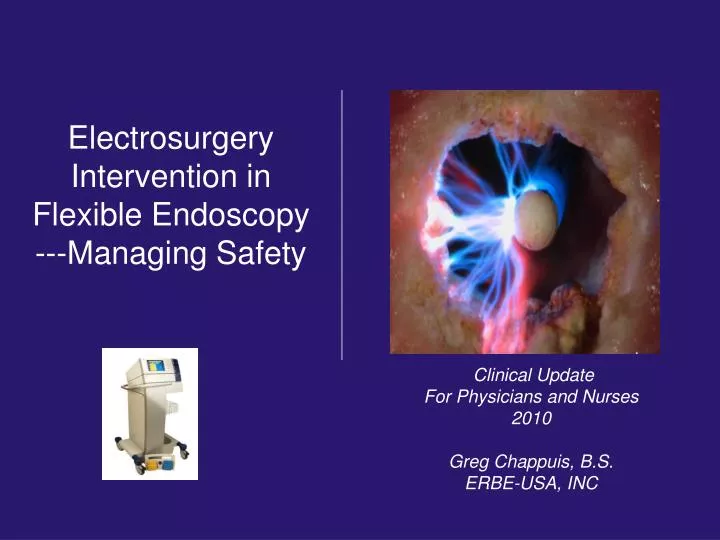
Electrosurgery Intervention in Flexible Endoscopy ---Managing Safety . Clinical Update For Physicians and Nurses 2010 Greg Chappuis, B.S. ERBE-USA, INC. HUMAN DIGESTIVE TRACT. Wall Thickness of the Small / Right Colon .

E N D
Electrosurgery Intervention in Flexible Endoscopy ---Managing Safety Clinical Update For Physicians and Nurses 2010 Greg Chappuis, B.S. ERBE-USA, INC
Wall Thickness of the Small / Right Colon The wall thickness of the right colon and small bowel is approximately 2mm which is less than three stacked pennies.
PEDUNCULATED POLYP • Basic pedunculated polyp.
SESSILE POLYPS • Sessile Polyp: Any polyp with a broad base.
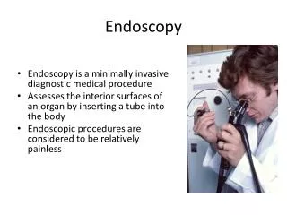
Hemostasis • Superficical bleeding
Many tools…many variables Submucosal fluid cushion? Type of generator? Braided – non braided snare? Cap assisted? Waveform?
> Formalized Education on Electrosurgery Survey of 400 Surgeons We are educated…but….
Basic Principles of Electrosurgery • Direct current = CauteryDirect current, which for example is generated by batteries, is not suitable for electrosurgical procedures because in addition to the desired thermal effect it also generates an undesirable electrolytic effect, producing acids and bases at the electrode poles. Danger of caustic burns !
Basic Principles of Electrosurgery • Alternating current Alternating current with frequencies which are normally used in every household (50-60 Hz) is not suitable for electrosurgical procedures because in addition to the desired thermal effect, these frequencies can produce an undesirable faradic effectresulting in neuromuscular stimulation. Muscle contractions !
Basic Principles of Electrosurgery Stimulating effect of alternating current on nerve and muscle cells as a function of frequency The frequency of the alternating current must be high enough to ensure that no neuromuscular stimulation is produced.Therefore only high-frequency alternating current with frequencies above 300.000 Hz (300kHz) is used in electrosurgery.
350,000 Hz ESU’s The Electrical FREQUENCY Spectrum (Why patients do not feel electrosurgery…) 54-880 MHz TV 100,000 Hz 60 Hz 550-1550 kHz Household Neuromuscular stimulation AM Radio
Circuit- flow of current from the • ESU to the active electrode, to the • patient, to the pad, and back to the ESU. The Clinical Circuit • Current – flow of electrons through • the electrical circuit. • Voltage - electrical force pushing • current around the circuit, through • varying degrees of tissue resistance. • Resistance (Impedance) - literally the • tissue being treated, which has varying • characteristics.
Clinical Translation of Ohm’s Law • Mathematically: Current = • Clinically: • Current increases as voltage increases • Current decreases as resistance increases • Remember: Current is the flow of electrons through a circuit in response to an applied electromotive force. Voltage Resistance
Always seeks ground. Two Basic Principles of Electricity • Always seeks the path of • least resistance.
Well vascularized area. • Shortest circuit possible. • Optimum – on flank. • Alternatives – • Thigh or Arm. • Avoid Buttock placement. • Remove pads carefully to • prevent shearing of skin. Dispersive Electrodes GI Endoscopy Pad Placement
Tissue Impedance Impedance Varies with Water Content of Tissue
There are Two Different TYPES of ESUs • CONSTANT POWER • Watts (power) setting is • chosen. • The Watts remain constant. • Voltage varies to maintain • Watts. • All tissue is treated with same • Wattage. • ValleyLab, Conmed,Endostat, • et al. • CONSTANT VOLTAGE • Wattage (power) maximum is • selected. • Voltage remains constant. • Microprocessors read tissue • response. • Watts (power) varies according to • tissue variables encountered at • the active electrode contactpoint. • ERBE
ESU Thermal Effects on Cells Temp Tissue Effect 104°F: Reversible cellular trauma 120°F: Irreversible cellular trauma 158°F: Coagulation (Desiccation) 212°F:Cutting 392°F: Carbonization
Basic Principles of ElectrosurgeryCutting Thermal Effects Cutting using high-frequency current • Essential: ! Sparking ! • Maximum current density • The extremely rapid vaporization of the intracellular liquid leads to the rupturing of the cell membrane • No mechanical force is required • Simultaneous hemostasis (adjustable) • (Vaporization)
Constant Power - Types of Electrical Waveforms Blend Blend is NOT a mixture of cut and coag. It is a modification of the duty cycle or coag “ON” time
Constant Voltage Voltage (EFFECT) EFFECT 1 EFFECT 2 EFFECT 3 EFFECT 4 • EFFECT - is how much voltage that is constantly being delivered to target tissue. As you increase EFFECT, hemostasis and thermal effect increases.
Types of Electrical Waveforms Cutting: Sinusoid (constant) • Voltage quickly raises water temperature in the cell to boiling point • Cell water turns to steam • Cell explodes, separating from adjoining cells • Cleavage plane is created = clinical “CUT”
Constant Voltage ERBE Regulation of Power Output The power output is dynamically regulated within the pre-set limits. It is independent of: • the cutting electrode • the direction of the cut • the tissue
Electrosurgical Cutting Waveform Endocut™ Proprietary waveform that involves a fractionated cutting mode characterized by alternating cutting and coagulation cycles.
Clinical Applications Sphincterotomy • Sphincterotomy Techniques • Pure or blended waveform controlled by pedal tapping. • Software controlled, fractionated cut / coag cycle with ‘pedal down’.
Efficacy of Using Endocut Akiho H, Sumida Y, Akahoshi K, Murata A, Ouchi J, Motomura Y, Toyomasu T, Kimura M, Kubokawa M, Matsumoto M, Endo S, Nakamura K. Safety advantage of endocut mode over endoscopic sphincterotomy for choledocholithiasis. World J Gastroenterol. 2006 Apr 7;12(13):2086-8. As noted in this study, efficacy of using endocut was shown in comparison To conventional blended cut mode for pancreatitis by reducing the hypermylasemia.
Efficacy of Using Endocut Perini RF, Sadurski R, Cotton PB, Patel RS, Hawes RH, Cunningham JT. Post-sphincterotomy bleeding after the introduction of microprocessor- controlled electrosurgery: does the new technology make the difference? GastrointestEndosc 2005; 61:53-57. The study revealed less endoscopic bleeding with the use of a microprocessor-controlled in comparison to conventional electrosurgery.
Clinical Benefits of CO2 Insufflation • Absorbed 150 times faster than room air – less • distention and intra and post operative pain. • Due to the rapid absorption, diminished • distention / pain post procedure occur • allowing the physician to quickly rule out • insufflation pain, in the event of pancreatitis or • perforation. Bretthauer M, et al. Carbon dioxide insufflations for more comfortable endoscopic retrograde cholangiopancreatopagraphy: a randomized, controlled, double-blind trial. Endoscopy 2007;39:58-64.
Clinical Applications Clinical Video: EndocutPolypectomy
Basic Principles of ElectrosurgeryCoagulation Thermal Effects Coagulation: • Hemostasisthrough ... • “Coagulation” of proteins... • Desiccation and shrinkage through the slow vaporization of cellular liquid and vascular occlusion • Devitalization • Tumors • Lesions • .........
Conventional Vs. AutomaticCoag Coag Conventional Automatic
Types of Electrical Waveforms Coag: Modulated (with resting points) • Current waveform with spikes of high voltage followed by rest periods • This allows the cellular proteins to slowly denature • Coagulation occurs
Electrosurgical Technique Hemostasisdepends on modulation and voltage Modulation - Coagulation Soft Forced Swift Dessicate Spray Fulgurate Keep in mind, you can have the wattage set at 60 watts per mode, but get very different tissue effects depending on the waveform and voltage associated with it.
Basic Principles of ElectrosurgeryBIPOLAR monopolar electrosurgery bipolar electrosurgery
Submucosal Injection Needle injection Needle-free submucosal injection Submucosal injection provides an additional cushion to protect the muscularis and also aids in dispersing electrosurgical current during electrosurgical procedures, including APC Norton ID, Wang LN, Levine SA, Bugart LJ, Hofmeister EK,Yacavo RF, et al. In vivo characterization of colonic thermalinjury caused by argon plasma coagulation. GastrointestEndosc 2002;55:631-6.
Submucosal Injection Submucosal needle-free injection
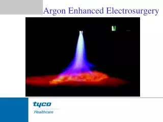
Argon Plasma Coagulation Electrode Argon Plasma Argon Gas Gas Flow in Probe Self-Limiting Desiccation Zone Mucosa When argon gas becomes electrically charged, it forms a plasma with a self-limiting desiccation zone
Non-contact application • As target tissue becomes coagulated, current automatically seeks new conductive tissue resulting in uniform hemostasis. • Smoke is reduced • Thinner eschar, more flexible • Limited penetration depth of approximately 3mm Argon Plasma Coagulation Advantages: Non-contact no sticking to tissue
APC GI Clinical Applications Gastroenterology Uses found in Clinical Literature • Radiation Induced Proctopathy • Watermelon Stomach (GAVE) • Treatment of Residual Adenomatous Tissue • Stent Shortening (e.g. migrated stents) • Strictures • Exophytic Benign or Malignant Tumors • Oozing from Vascular Lesions(e.g. Angiodysplasias, • Arteriovenous Malformations(AVMs), Telangiectasias)
Argon Plasma Coagulation Gastroenterology Uses found in Clinical Literature References: • “The role of endoscopy in ampullary and duodenal adenomas”. Gastrointestinal Endoscopy; 2006: Vol. 64, No 6. • Brooker, J. Treatment with APC reduces recurrence after piecemeal resection of large sessile colonic polyps: a randomized trial and recommendations. Gastrointestinal Endoscopy, 2002. • Buyukberber, Mehmet. APC in the treatment of hemorrhagic radiation proctitis. Turk J Gastroenterol, 2005. • Dulai, Gareth. Treatment of Water Melon Stomach. Current Treatment Options in Gastroenterology, 2006. • Eickhoff, A, et al. Prospective nonrandomized comparison of two modes of argon beamer (APC) tumor desobstruction: effectiveness of the new pulsed APC versus forced APC. Endoscopy 2007: 39: 637-642. Ferreira, L, et al. Post-Sphincterotomy Bleeding: Who, What, When, and How. American Journal of Gastroenterology. 2007. • Eickhoff, A, et al. Pain sensation and neuromuscular stimulation during argon plasma coagulation in gastrointestinal endoscopy. Surg Endosc. 2007. • Fujishiro, M. Safety of Argon Plasma Coagulation for Hemostasis During Endoscopic Mucosal Resection. Surg Laparosc Endosc Percutan Tech; 2006. • Fukami, N. Endoscopic treatment of large sessile and flat colorectal lesions. Current Opinions in Gastroenterology. 2006:22:54-59. • Fukatsu, H, et al. Evaluation of needle-knife precut papillotomy after unsuccessful biliary cannulation, especially with regard to postoperative anatomic factors. Surg Endosc. 2008;22:717-23. • Garcia, A, et al. Safety and efficacy of argon plasma coagulator ablation therapy for flat colorectal adenomas. Rev Esp Enferm Dig. 2004:96:315-321. • Herrera S, et al. The beneficial effects of argon plasma coagulation in the management of different types of gastric vascular ectasia lesions in patients admitted for GI hemorrhage. Gastrointestinal Endoscopy 2008. • Horiuchi, A, et al. Effect of precut sphincterotomy on biliary cannulation based on the characteristics of the major duodenal papilla. Clin Gastroenterol Hepatol. 2007;5:1113-8. • Ifadhli A, et al. Efficacy of argon plasma coagulation compared with topical formalin application for chronic radiation proctopathy. Can J Gastroenterol 2008;22:129-132. • Kitamura, Tadashi. Argon plasma coagulation for early gastric cancer: technique and outcome. Gastrointestinal Endoscopy, 2006. • Kwan, V. APC in the Management of Symptomatic GI Vascular Lesions. American Journal of Gastroenterology. 2006. • Lecleire, S, et al. Bleeding gastric vascular ectasia treated by argon plasma coagulation: a comparison between patients with and without cirrhosis. Gastrointestinal Endoscopy. 2008:67.
Argon Plasma Coagulation Gastroenterology Uses found in Clinical Literature Cont. • References: • 17. Manner, H, et al. Safety and efficacy of a new high power argon plasma coagulation system (hp-APC) in lesions of the upper gastrointestinal tract. Digestive and Liver Disease. 2006. • 18. Norton, I, et al. A Randomized Trial of Endoscopic Biliary Sphincterotomy Using Pure-Cut Versus Combined Cut and Coagulation Waveforms. Clinical Gastroenterology and Hepatology. 2005; 3:1029-1033. • 19. Norton, I, et al. Efficacy of colonic submucosal saline solution injection for the reduction of iatrogenic thermal injury. Gastrointestinal Endoscopy. 2002:Vol 56, No 1. • 20. Olmos, Jorge. APC for prevention of recurrent bleeding from GI angiodysplasias. Gastrointestinal Endoscopy, 2004. • 21. Ortner, M, et al. Endoscopic Interventions for Preneoplastic and Neoplastic Lesions: Mucosectomy, Argon Plasma Coagulation, and Photodynamic Therapy. Digestive Diseases. 2002;20 :167-172. • 22. Perini, Rafael. Post-sphincterotomy bleeding after microprocessor-controlled electrosurgery. Gastrointestinal Endoscopy. 2005. • 23. Regula, J. Argon Plasma Coagulation after Piecemeal Polypectomy of Sessile Colorectal Adenomas: Long-Term Follow-Up Study. Endoscopy, 2003. • 24. Repici, A. Endoscopic polypectomy: techniques, complications and follow-up. Tech Coloproctol. 2004; 8: S283-S290. • 25. Rerknimitr, R. Trimming a Metallic Biliary Stent Using an Argon Plasma Coagulator. Cardio Vascular and Interventional Radiology, 2006. • 26. Ross, A. Flat and Depressed Neoplasms of the Colon in the Western World. American Journal of Gastroenterology. 2006. • 27. Schubert, D. Endoscopic treatment of benign gastrointestinal anastomotic strictures using argon plasma coagulation in combination with diathermy. Surg Endosc; 2003:17:1579-1582. • 28. Soctikno, R, et al. Prevalence of Nonpolypoid (Flat and Depressed) Colorectal Neoplasms in Asymptomatic and Symptomatic Adults. JAMA. 2008: Vol 299, No 9. • 29. Vargo, John. Clinical Applications of APC. Gastrointestinal Endoscopy, 2004. • 30. Zlatanic, J, et al. Large sessile colonic adenomas: use of argon plasma coagulator to supplement piecemeal snare polypectomy. Gastrointestinal Endoscopy; 1999: Vol. 49, No. 6.
Argon Plasma Coagulation Pulsed 2 APC GAVE
Argon Plasma Coagulation The extent of the thermal effect of APC on tissue depends on several factors:
Argon Plasma Coagulation Another important factor involving thermal effect is the mode chosen • APC has evolved through specialized modes with more controllable thermal effect: • Pulsed 1 APC: pulses one time per • second, used for focused coagulation • Pulsed 2 APC: pulses 16 times per • second, used for wide spread • coagulation • Forced APC: Constant beam, often • used for devitilization of tissue (Original • APC for GI – ERBE APC 300 circa 1992 – • used Forced or constant beam only)
Argon Plasma Coagulation Modes • Precise APC: • The Precise mode creates a more • superficial coagulation effect using • a low-energy output, suitable for • temperature sensitive, thin-walled areas • Due to its potential to auto-regulate the • beam by increasing and decreasing • intensity with probe movement (i.e • distance in relation to target tissue), • thermal effect is more homogenous Regula J, Wronska E, et al. Vascular lesions of the gastrointestinal tract. Best Practice and Research Clinical Gastroenterology 2008; 22: 313-328