Download
1 / 80
820 likes | 1.54k Views
Sources. Flow Cytometry and Sorting, 2nd ed. (M.R. Melamed, T. Lindmo, M.L. Mendelsohn, eds.), Wiley-Liss, New York, 1990 - referred to here as MLMFlow Cytometry: Instrumentation and Data Analysis (M.A. Van Dilla, P.N. Dean, O.D. Laerum, M.R. Melamed, eds.), Academic Press, London, 1985 - VDLM. Sources (continued).

E N D
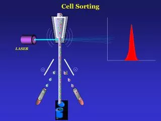
1. Flow Cytometry and Cell Sorting
Adapted by Albert D. Donnenberg, Ph.D. from:
�Fluorescence Spectroscopy in Biological Research�
Robert F. Murphy, Ph.D. Carnegie Mellon University
2. Sources Flow Cytometry and Sorting, 2nd ed. (M.R. Melamed, T. Lindmo, M.L. Mendelsohn, eds.), Wiley-Liss, New York, 1990 - referred to here as MLM
Flow Cytometry: Instrumentation and Data Analysis (M.A. Van Dilla, P.N. Dean, O.D. Laerum, M.R. Melamed, eds.), Academic Press, London, 1985 - VDLM
3. Sources (continued)
4. Definitions Flow Cytometry
Measuring properties of cells in flow
Flow Sorting
Sorting (separating) cells based on properties measured in flow
Also called Fluorescence-Activated Cell Sorting (FACS)
5. Basics of Flow Cytometry
6. Fluidics Need to have cells in suspension flow in single file through an illuminated volume
In most instruments, accomplished by injecting sample into a sheath fluid as it passes through a small (50-300 �m) orifice
8. Fluidics When conditions are right, sample fluid flows in a central core that does not mix with the sheath fluid
This is termed Laminar flow
9. Whether flow will be laminar can be determined from the Reynolds number
When Re < 2300, flow is always laminar
When Re > 2300, flow can be turbulent Fluidics - Laminar Flow
10. Fluidics The introduction of a large volume into a small volume in such a way that it becomes �focused� along an axis is called Hydrodynamic Focusing
11. Fluidics
12. Fluidics
13. Fluidics
14. Fluidics How do we accomplish sample injection and regulate sample flow rate?
Differential pressure
Volumetric injection
15. Fluidics - Differential Pressure System Use air (or other gas) to pressurize sample and sheath containers
Use pressure regulators to control pressure on each container separately
16. Fluidics - Differential Pressure System Sheath pressure will set the sheath volume flow rate (assuming sample flow is negligible)
Difference in pressure between sample and sheath will control sample volume flow rate
Control is not absolute - changes in friction cause changes in sample volume flow rate
17. Fluidics - Differential Pressure System
18. Fluidics - Volumetric Injection System Use air (or other gas) pressure to set sheath volume flow rate
Use syringe pump (motor connected to piston of syringe) to inject sample
Sample volume flow rate can be changed by changing speed of motor
Control is absolute (under normal conditions)
19. Volumetric Injection System
20. Fluidics - Particle Orientation and Deformation As cells are hydrodynamically focused, they experience shear stresses on different points on their surfaces (an in different locations in the stream)
These cause cells to orient with their long axis (if any) along the axis of flow
The shear stresses can also cause cells to deform (e.g., become more cigar-shaped)
21. Particle Orientation and Deformation
22. Fluidics - Flow Chambers The flow chamber
Defines the axis and dimensions of sheath and sample flow
Defines the point of optimal hydrodynamic focusing
Can also serve as the interrogation point (the illumination volume)
23. Fluidics - Flow Chambers Four basic flow chamber types
Jet-in-air
best for sorting, inferior optical properties
Flow-through cuvette
excellent optical properties, can be used for sorting
Closed cross flow
best optical properties, can�t sort
Open flow across surface
best optical properties, can�t sort
24. Fluidics - Flow Chambers
25. Fluidics - Flow Chambers
26. Fluidics - Flow Chambers
27. Optics Need to have a light source focused on the same point where cells have been focused (the illumination volume)
Two types of light sources
Lasers
Arc-lamps
28. Optics - Light Sources Lasers
can provide a single wavelength of light (a laser line) or (more rarely) a mixture of wavelengths
can provide from milliwatts to watts of light
can be inexpensive, air-cooled units or expensive, water-cooled units
provide coherent light
29. Optics - Light Sources Arc-lamps
provide mixture of wavelengths that must be filtered to select desired wavelengths
provide milliwatts of light
inexpensive, air-cooled units
provide incoherent light
30. Optics - Optical Channels An optical channel is a path that light can follow from the illuminated volume to a detector
Optical elements provide separation of channels and wavelength selection
31. Optics - Forward Scatter Channel When a laser light source is used, the amount of light scattered in the forward direction (along the same axis that the laser light is traveling) is detected in the forward scatter channel
The intensity of forward scatter is most influenced by the size of cells (or other particles)
33. Optics - Side Scatter Channel When a laser light source is used, the amount of light scattered to the side (perpendicular to the axis that the laser light is traveling) is detected in the side or 90o scatter channel
The intensity of side scatter is most influenced by the shape and optical homogeneity of cells
35. Optics - Light Scatter Forward scatter tends to be more sensitive to surface properties of particles (e.g., cell ruffling) than side scatter
can be used to distinguish live from dead cells
Side scatter tends to be more sensitive to inclusions within cells than forward scatter
can be used to distinguish granulated cells from non-granulated cells
36. Optics - Fluorescence Channels The fluorescence emitted by each fluorochrome is usually detected in a unique fluorescence channel
The specificity of detection is controlled by the wavelength selectivity of optical filters and mirrors
38. Optics - Filter Properties Optical filters are constructed from materials that absorb certain wavelengths (while transmitting others)
Transitions between absorbance and transmission are not perfect; the sharpness can be specified during filter design
39. Optics - Filter Properties Filters must have very sharp cutons and cutoffs since scattered laser light is several orders of magnitude greater than emitted fluorescence
Filters are designed to reject light to specific tolerances (e.g., reject 488 nm light at 10-6 level: only 0.0001% of incident light at 488 nm gets through)
40. Optics - Filter Properties Long pass filters transmit wavelengths above a cut-on wavelength
Short pass filters transmit wavelengths below a cut-off wavelength
Band pass filters transmit wavelengths in a narrow range around a specified wavelength
Band width can be specified
41. Standard Long Pass Filters
43. Optics - Filter Properties When a filter is placed at a 45o angle to a light source, light which would have been transmitted by that filter is still transmitted but light that would have been blocked is reflected (at a 90o angle)
Used this way, a filter is called a dichroic filter or dichroic mirror
44. Dichroic Filter/Mirror
45. Optics - Filter Layout To simultaneously measure more than one scatter or fluorescence from each cell, we typically use multiple channels (multiple detectors)
Design of multiple channel layout must consider
spectral properties of fluorochromes being used
proper order of filters and mirrors
48. (Overhead 10) Channel Layout for Arc Lamp-based Flow Cytometry
49. Optics - Detectors Two common detector types
Photodiode
used for strong signals when saturation is a potential problem (e.g., forward scatter detector)
Photomultiplier tube (PMT)
more sensitive than photodiode but can be destroyed by exposure to too much light
50. Wavelength Dependence of Photomultipliers
51. Electronics Processing of signals from detectors
Preamplification
Strengthen signals so that they can travel from remote detectors to central electronics
Amplification
Adjust signal intensity
Linear or Logarithmic
Log transformation can also be performed after digitization using a look-up table
52. Comparison of linear and logarithmic amplification
53. Electronics Processing of signals from detectors
Generation of Integral or Pulse Width
Gated Peak-Sense-And-Hold
Timing Adjustment
Necessary for multiparameter systems
Analog-Digital Conversion
54. Signal Processing
55. Data Acquisition Each measurement from each detector is referred to as a �parameter�
Data are acquired as a �list� of the values for each �parameter� (variable) for each �event� (cell)
56. Listmode Data Acquisition
57. Single parameter histograms
58. Bivariate Histograms
59. Gating
60. Basics of Flow Sorting Droplet formation
Timing
Coincidence - Purity and Efficiency
62. Droplet formation
63. Timing
64. Coincidence - Purity As droplets form, they can contain wanted cells as well as unwanted cells. If all droplets containing a wanted cell are sorted (regardless of whether they also contain unwanted cells), the purity of the sorted sample will be reduced.
65. Coincidence - Purity The purity can be improved by checking for coincidence events and not sorting any wanted cell that occurs too close to an unwanted cell.
This causes an increase in purity but a reduction in sorting efficiency.
66. Coincidence - Efficiency
67. Cell Cycle Analysis One of the earliest applications of flow cytometry was the analysis of cell cycle position by quantifying cellular DNA.
Flow cytometry is still the method of choice for fast, accurate determination of cell cycle distributions.
68. Univariate Cell Cycle Methods In the simplest method, cellular DNA is detected using a fluorescent dye that binds preferentially to DNA.
Propidium iodide is most commonly used. It undergoes a dramatic increase in fluorescence upon binding DNA. It requires permeabilization of the plasma membrane.
Hoechst 33342 can be used where labeling of unpermeabilized (live) cells is desired.
69. Univariate Cell Cycle Methods When the amount of DNA per cell is measured on a sample from an asynchronously growing cell culture, cells with various amounts of DNA from the 2N (G0/G1) amount to the 4N (G2/M) amount are observed. A histogram reveals the fraction of cells in the various cell cycle phases.
70. Normal Cell Cycle
71. DNA Analysis
72. Cell cycle progression of synchronized cells
73. DNA Analysis
74. Bivariate Cell Cycle Analysis To aid in the detection of cells in S-phase, a brief pulse of a marked nucleotide can be used. The most common such nucleotide is bromodeoxyuridine (BrdU) which is incorporated into DNA in place of thymidine. The incorporated BrdU can be detected with an antibody, identifying those cells that synthesized DNA during the pulse.
75. Detection of incorporated BrdU
77. Chromosome Analysis and Sorting Individual chromosomes can be analyzed in flow after appropriate preservation and isolation. The most common method is to use two different DNA dyes, one (Hoechst 33258) that binds preferentially to AT-rich DNA and one (chromomycin A3) that binds preferentially to GC-rich DNA.
78. Two-color chromosome analysis
80. Immunofluorescence Analysis A major application of flow cytometry is the analysis (and sorting) of subsets of blood cells using surface markers.
A useful feature is that the major blood cell types show distinct forward and side scatter profiles.