Download

1 / 12

E N D
Osteogen Osteogenesis D esis Distraction istraction Part (1) Part (1) Prepared by: Prepared by: Mohammed Alruby Mohammed Alruby Part 1 Osteogenesis Distraction Osteogenesis Distraction Part 1 Dr. Mohammed Alruby Dr. Mohammed Alruby
2 = Definition = Historical development of craniofacial distraction. = Distraction device classification. = Biological aspect of osteogensis distraction. = Distraction histogensis: Effect on muscle Effect on the peripheral nerves. Effect on TMJ. Effect on periodontal ligament. Effect on gingival tissues. = Biomechanical effect of distraction device orientation during mandibular lengthening and widening: In transverse plane In Sagittal plane Dr. Mohammed Alruby Dr. Mohammed Alruby O Osteogenesis steogenesis Distraction Part Distraction Part 1 1
3 Distraction Osteogenesis Definition: Is a biologic process of new bone formation between the surfaces of bone segments that are gradually separated by incremental traction? This traction force, in turn- generate tension within the tissues that connect the bone segments, which stimulate new bone formation parallel to the vector of distraction. Importantly, distraction force applied to bone also create tension in the surrounding soft tissues, initiating a sequence of adaptive changes termed distraction histogensis. Under the influence of tensional stress produced by gradual bone distraction, active histogensis occurs in different tissues, including gingiva, blood vessels, ligaments, cartilage, muscles and nerves. These adaptive changes in the soft tissues allow larger skeletal movements while minimizing the relapse seen in acute orthopedic correction History: - Although distraction osteogensis offer novel solutions for correction of sever maxillary and manbibular deformities, the application of tensile and compressive forces to bones of craniofacial skeleton is not anew concept. Since the 1800, mechanical forces were used to separate the bones of maxilla at the midpalatal suture. More-over, depending on the maturational status of the patient, different distraction like palatal expansion procedures have been applied. The concept and the technique of distraction osteogensis were first described by- Codvilla (1905) who tried distraction of long bone that modified in 1951 by Gavrill Ilizarov that design a new apparatus for bone fixation ==Historical development of craniofacial distraction: ** Dentofacial traction: In 1728 Fauchard describe the use of expansion arch that apply tensile and compressive force, when ligating an ideally shaped metal plate to the teeth, this form of traction was limited to tooth movement only and had limited effect on the shape of the bone. In 1859 Wescott the first reported the placement of the mechanical force to the bone of maxilla, a year later Angell performed a similar procedure, using a differentially threaded jackscrew connected across the palate to both bicuspid on one side and the second bicuspid on the other side In 1893 Goddard further standardized the palatal expansion protocol. ** Craniofacial osteotomies: The first surgical procedure for the correction of the craniofacial deformity was reported in 1848 by Hullihen that treat the pro-gnathic mandible, after that Blair demonstrate the use of bilateral horizontal ramus osteotomy to advance the mandible for treatment of case of class II. In an attempt to increase the contact surface area between divided bone segment and provide greater stability of bone fixation as Eieslberg and Pehrgadd that developed step like sliding osteotomies for lengthening or widening the mandible In 1927 Rosethal performed the first mandibular osteo-distraction procedure by using an intraoral tooth born appliance that gradually activated over a period of 1 month. Ten years later, Kazanjian performed mandibular osteo-distraction by using gradual incremental traction instead of acute advancement. In 1948 Crawford applied gradual incremental traction to the fractured Dr. Mohammed Alruby Dr. Mohammed Alruby O Osteogenesis steogenesis Distraction Part Distraction Part 1 1
4 callus of the mandible, in 1959 Kole, describe a method for surgically correcting an anterior openbite due to maxillary anterior deformity and in the same year Kole also developed the predecessor of rapid canine distraction method by using of Corticotomy technique to Assis the moving teeth as individual or as group with orthodontic mechanics. In the late of the same year Kole was also the first to demonstrate the combination of orthodontic and surgical technique for rapid expansion of the palate on adults. **Mandibular distraction: In 1989 Mccarthy and colleagues were the first to clinically apply extraoral distraction in the mandible. In 1990 Gurerrero developing his midsymphyseal mandibualr widening technique using an intraoral tooth born device. In 1995 Molina and Ortize- Monesterio simplified the methods established by Mccarthy **Maxillary and midface distraction: The first study demonstrating the possibility of maxillary distraction was reported by Rachmiel in 1993, in 1995 Block and associate demonstrate maxillary advancement using tooth born distraction device in dog and in the same year Polley and others report the first clinical application of midface distraction. ** Alveolar ridge distraction: In 1996, Chin and Toth reported the first clinical application of mandibular alveolar distraction osteogensis, this technique is indicated in case of: - Developmental anomalies such as: cleft palate. - Maxillofacial truma that involve damage of the teeth and associated jaw structure. - Periodontal disease that lead to bone and tooth loss from the alveolar process ** Distraction device classification: = According to location: 1-Extraoral device: that are attached to the bone by percutaneous pins Connected externally to fixation clamps that divided into: unidirectional Bi-directional Multidirectional 2- Internal device: are located either subcutaneously or within the oral cavity (Intraorally), the intraoral device can be placed above and called extra mucosal or below and called submucosal. = According to anchorage: -Bone born -Tooth born -Both: hybrid Biological aspect of Osteogenesis distraction - Distraction osteogensis is a unique process of new bone formation between the surface of the bone segments that are gradually separated by incremental traction. Depending on the anatomic site where the traction is induced distraction osteogensis technique are divided into 1- Callotasis: which means distraction of the fractured callus. 2- physeal distraction: which distraction of the bone growth plates. Dr. Mohammed Alruby Dr. Mohammed Alruby O Osteogenesis steogenesis Distraction Part Distraction Part 1 1
5 A-distraction epiphysiolysis: involve a relatively rapid rate of bone segment separation usually ranging from 1.0 to 1.5 mm. Per day, the rapidly increased tension at the growth plate produce a fracture of the physis, the subsequent gradual separation of the bone lead to replacement of the growth plate cartilage by trabecular bone. B- chondrodiatasis: utilize a very slow rate of bone segment separation (less than 0.5mm / day) this allow stretching of growth plate without fracture. - Callotiasis begins with the development of a reparative callus similar to that observed during bone repair or fracture healing. New bone formation is initiated when a traction force is applied to the bone segments, thereby interrupting the process of fracture healing and placing the callus under tension. As the callus stretched, new bone is regenerated parallel to the direction of traction. During this stretching, the soft callus is maintained at the center of distraction gap while routine fracture healing occurs at the periphery of the regenerate. - Clinically, distraction osteogensis consists of five sequential periods: 1- Osteotomy: divides a bone into two segments, resulting in loss of continuity and mechanical integrity. 2- Latency period: is the period from bone division to the onset of traction, this represent time allowed for reparative callus formation. Initially as a result of vascular disruption, hematoma is forms between and around the bone segment, hematoma is converted to clot and bony necrosis at the end of the fractured segments. Lasts from 1 to 3 days, at which time the clot is replaced with granulation tissue consisting of inflammatory cell, fibroblast, collagen and invading capillaries. Following that is formation of soft callus and this period is marked by continuo ingrowth of capillaries, during this stage the granulation tissue is converted to fibrous tissue by fibroblast. Cartilage also replaced the granulation tissue, this occur more toward the periphery of the inter-segmentary gap than the central region. 3- Distraction period: characterized by the application of traction to osteotomized bone segments that are gradually pulled apart, resulting in formation of new bony tissue within the progressively increasing intersegmental gap. Through the application of tension stress to the inter-segmentary tissue of the soft callus, the tension stress that develops in the gradually stretched tissue stimulate changes at cellular and subcellular levels, these changes can be characterized as growth stimulating effect and a shape forming effect. Between the third and seventh days of distraction capillaries grow into the fibrous tissues, thereby extending the vascular network not only toward the center of the gap but also toward the medullary canal of both adjacent bone segments. During the second week of distraction, primary trabeculae begin to form. The osteoblasts located among the collagen fibers lay down osteoid tissue on these collagen fibers, by the end of the second week the osteoid begins to mineralize. 4- Consolidation period: is that time between cessation of traction forces and removal of the distraction device. The period represents the time for complete mineralization of the distraction degenerate. Dr. Mohammed Alruby Dr. Mohammed Alruby O Osteogenesis steogenesis Distraction Part Distraction Part 1 1
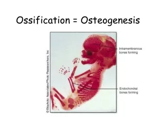
6 5- Remodeling period: is the period from the application of full functional loading to the complete remodeling of the newly formed bone. It takes a year or more before the structure of newly formed bony tissue is comparable to that of the preexisting bone. N.B: the mechanism of new bone formation during distraction osteogensis is dependent on both the blood supply and rate of distraction. The most appropriate rate of mandibular distraction is between 0.5 and 1.0 mm / day. **Tooth movement into regenerate bone formed: Several experimental studies were made to describe the tooth movement for the distracted area (area of regenerate bone) at the beginning of distraction (group I), at the end of distraction (group 2) and at the consolidation period (group 3). The studies indicated that the movement of the tooth in-group 2 is more accepted movement and more safes during and after movement and there are several factors that affect the tooth movement: 1- systemic: bone quality calcium level General health blood supply tooth born related factors: =Size and shape of the tooth root. =The quality of the surrounding bones =Amount of bone between osteotomy and tooth root. mechanical factors: The type of tooth movement. The stretched dentoalveolar and gingival fibers during distraction that applying forces to the tooth. **Implant and distraction osteogensis: The technique of distraction osteogensis allows the creation of significantly more alveolar bone than commonly achieved with grafts, which may enhance the overall prosthetic rehabilitation as well as esthetics. So several studies were made to analyze the osteo-integration of endosseous implant placed into the regenerated bone area, the radiographic evaluation of these studies indicated that: Immediately after implant placement no bone was observed around the implant but after four weeks the density of bone trabeculae had increased, twelve weeks after implant placement the distraction regenerate was totally mineralized and no radiolucent area was observed at the implant area. By histological evaluation: Twelve week after implant placement newly formed lamellar bone observed around the implant, however only few areas with direct bone / implant contact were seen, after 24 weeks for implant placement, there are abundant newly formed mature lamellar bone around the implant. ** Histology of bone 1 year after distraction: The mechanism of bone formation during osteo-distraction is controversial, Karp in 1992 reported that the bone formed intramembranous method but Komuro in 1994 demonstrated that the bone was formed by both endochondral and intramembranous bone formation especially in the mandibular distraction. This supported by the presence of fibrocartilage island within 1- 2- - Dr. Mohammed Alruby Dr. Mohammed Alruby O Osteogenesis steogenesis Distraction Part Distraction Part 1 1
7 human mandibular regenerate bone, all the histologic evaluation of the regenerated area indicated that the bone reach quality after 1 year from distraction. Distraction histogenesis: Lengthening of the soft tissue is an important part of the distraction osteogensis process. Ideally, the bone segment and the associated soft tissue should lengthen proportionally. However, bone lengthening is possible because the bone is surgically divided into multiple segments, whereas the soft tissues are stretched without surgical separation. Because of this, different biologic mechanisms are involved in the soft tissue response to gradual stretching. This process is termed distraction histogenesis. Distraction histogensis is a biologic process of soft tissue adaptation to gradual stretching. This process is initiated by the tension created when applying distraction force to the bone segments. Under the influence of tension stress, active adaptation occurs in all of the soft tissues. Depending on the elasticity and capacity of regeneration, the various soft tissues respond differently to gradual stretching. Nonetheless, two predominant mechanisms of soft tissue adaptation occur during distraction histogenesis: (1) soft tissue regeneration following disruptive and degenerative changes and (2) neo histogenesis as a result of generalized cellular proliferation and growth. Effect on muscles: The structural and functional unite of the muscles is the muscle fibers that covered by thick membrane called sarcolemma and surrounded by connective tissue called Endomysium. The muscle fiber coalesces together to form muscle bundles; each bundles is covered by connective tissue called perimysium. -The muscle bundles coalesce together to form the whole muscle which covered by connective tissue fascia called epimysium, these connective tissue contain blood vessels, nerves and lymph vessels. The sarcomer is the smallest functional unite of muscle contraction, it has been shown that the force developed by muscle during isometric contraction is dependent on sarcomere length. - In order to preserve or restore muscle function, the length of sarcomeres must be returned to the optimal range and this depend on one of these mechanisms: the sarcomer length decrease during skeletal lengthening due to decrease the distance between the muscle origin and insertion point. Optimizing the sarcomer length in stretched muscle fiber is by rearranging the muscle fiber relative to the axis of force generation. - The length of the sarcomer could also be returned to the optimal range by addition of new sarcomeres within the stretched muscle fibers. The result from several studies for histologic evaluation of the geniohyoid muscle indicated that certain range of adaptive changes in response to 10 mm. of bilateral mandibular lengthening- some changes such as local atrophy of myofibers, focal necrosis, were unspecific and fairly limited in extent. Concurrently, there was evidence of muscle fiber regeneration in response to muscle traction. Dr. Mohammed Alruby Dr. Mohammed Alruby O Osteogenesis steogenesis Distraction Part Distraction Part 1 1
8 ** Bone – muscle lengthening ratio: Several studies demonstrate that muscles take smaller length increase than the bone, as Day and co – workers studies, demonstrate 6% increase of muscles with 10% tabial lengthening. **Does muscle grow or stretch: A number of studies were designed to address the fundamental question of whether gradual distraction promotes muscle growth or whether muscle fibers are stretched. - Matan and co- authors described the changes in sarcomer length, which increased immediately after bone elongation, then decreased gradually and return to pre-distraction values with concurrent increase in number of sarcomeres - histological studies also support the hypothesis of muscles growth under the influence of tension stress. These studies have demonstrated that muscle neogenesis does occur during gradual distraction and is thought to progress from myoblast proliferation to myoblast fusion into myotubes. - Other studies based on: progressive bone distraction progressively stretches the muscles, resulting in repetitive micro-lesions and focal necrosis of individual muscle fibers, macrophages immediately migrate to the disrupted fibers to engulf the muscle fibers. Damage of the basal lamina stimulate the release of stellite cells, which play an integral role in regeneration and growth of skeletal muscles. Release and activation of stellite cells coincide with massive proliferation of myoplast, the myoplast then fuse into myotubes, which after synthesis of contractile elements give rise to the new muscle fibers growing independently or incorporating with the ends of existing muscle fibers. - With regarding to the rate and rhythm of distraction, Illizarove and other investigators demonstrated excellent bone regenerate formation and favorable soft tissue response with the rate of distraction 1 mm./ day. - Other finding suggest a more fractionated rhythm of distraction promotes more effective myogenesis, this is in arrangement with a report of Mizumoto and co-authors, they found that an increased frequency of distraction resulted in higher DNA synthesis thereby providing better muscle growth and regeneration during lengthening. **Safety amount of distraction: It noted that limb lengthening up to 10% to 12% poses no damage to the muscle and other associated soft tissue, with up to 10% lengthening the muscle may stretch to accommodate the increased length. - With greater lengthening, the muscles undergo changes including myo-fiber atrophy, degeneration. Several studies to compare the results for increasing the length more than 20% indicated irreversible muscle damage. Effect of gradual traction on peripheral nerves: The nervous system is composed of an intercommunicating network of specialized cells called neuron, which are characterized by a cell body, along process (axon), numerous short process (dendrites), and specialized cell functions (synapse). The neuron is the structural and functional units of the nervous system. The overall studies results demonstrate that distraction osteogensis might produce significant abnormalities on inferior alveolar nerve (IAN) function. These abnormalities primarily resulted from the surgical procedure (either osteotomy or fixation screw placement). Dr. Mohammed Alruby Dr. Mohammed Alruby O Osteogenesis steogenesis Distraction Part Distraction Part 1 1
9 - The osteotomy is one of the most critical procedure for IAN integrity and therefore has to be performed with careful attention to the neurovascular anatomy and its individual variation, several mandibular osteotomy techniques have been developed to minimize trauma to the neurovascular bundles and enhance new bone formation during subsequent distraction. Some authors place full thickness cortical drill holes along the osteotomy line and then connect them with reciprocating or oscillating saw, and others divide lateral, superior and inferior aspect of the mandible leaving the medial cortex intact N.B: IAN injury may also be related to insertion the fixation pins or screws for distraction device anchorage, Extraoral devices with transcutaneous endosseous pins have been associated with higher risk of direct IAN injury. **Adaptation of peripheral nerves to gradual incremental traction Several experimental studies have demonstrated that peripheral nerve trunk are highly resistant to stretching. Wall and co-workers reported that during acute tabial rabbit nerve stretching, significant nerve conduction changes were first observed at 6% strain. At 12% strain, complete nerve conduction block was documented. ** Amount and rate of distraction: Illiazarov demonstrate progressive degenerative changes of nerve fibers for 50% limb lengthening, the first 5% of lengthening was mainly characterized by early degenerative changes of myelinated nerve fibers with segmental constriction of nerve axons and shwann cells. It was postulated by Illizarov that 1.0 mm rate of daily distraction performed in a high fractionated rhythm creates the most optimal environment for both regenerate bone formation and surrounding soft tissue adaptation Effect of gradual traction on gingival tissue From several studies on the gingival tissue due to distraction osteogensis, the results indicated that gingival tissue responds favorably to gradual stretching during osteo-distraction. It initially undergoes mild inflammation and reactive changes during distraction for few weeks of consolidation, followed by reparative changes with neo-histogenesis to restore the structural and functional integrity of the masticatory mucosa during the second through the eight week of consolidation. Effect of gradual traction on the periodontal ligament The PDL is the connective tissue that attaches the tooth root to the alveolar process, although the PDL thickness ranges from 0.15 to 0.38 mm, it is fairly consistent around the tooth. The PDL is composed of collagen fibrils that protect the tooth against forces from multiple direction. The mechanism of PDL adaptation to gradual traction during craniofacial distraction osteogensis is similar to that for orthodontic tooth movements, by starting compression and tension stimulate adaptive mechanisms as resorption, cemento-genesis and thereby restoring the equilibrium in length and tension of PDL. The sequence of adaptive changes is affected by the placement of distraction forces, whether to the tooth or to bone segment. Tooth born distraction device do not transmit all distraction forces to the bone segments due to the elasticity of the surrounding soft tissue and stretching of PDL, this often results in greater dental movement than skeletal movement and therefore should be taken into account during preoperative planning when calculating distraction parameters such as duration of the - - - Dr. Mohammed Alruby Dr. Mohammed Alruby O Osteogenesis steogenesis Distraction Part Distraction Part 1 1
10 distraction period and distraction rate. Although bone borne distraction devices produce proportional skeletal movements, dental relapse and tipping of the adjacent teeth may often occur. Therefore, preventive measures such as using full-sized arch wires completely ligated into place or retaining teeth during distraction and consolidation are probably indicated. Effect on TMJ: Since the mandible and TMJ are considered a functional unite, so the mechanical loading of the osteotomized mandible may have effect on joint position and physiology. * Biologically: the TMJ is a synovial joint like other joint in the body is composed of articular cartilage – synovial fluids – synovial membrane. The synovial fluids cover the articular surface, and essential for joint lubrication and nutrition. Structural alteration in the synovial membrane may cause changes in the content of the synovial fluids, resulting in altered cartilage metabolism. From the biomechanical point of view, there are several factors that influencing TMJ during distraction osteogensis as: 1-Movement within the architecture of the joint. 2-Restriction imposed by the soft tissue envelope. 3-The load characteristic particularly of the articular surface and the applied pressure. * Physiologically: articular cartilage provides a nearly frictionless surface for transmission and distribution of joint loads exhibiting little or no wear over decades of use. The maintenance of the articular cartilage due to the balance of the anabolic and catabolic activities of chondrocyte cell population. The results from several studies about the changes on TMJ, revealed that: 1-Permanent degenerative alteration in the TMJ leading to a clinical impairment of joint function. 2- Initial disturbance may be associated with adaptive joint remodeling and subsequent growth stimulation. 3- The joint may functionally adapt to the changing mechanical environment by altering structural integrity. According to these results some author support distraction protocol without extensive mechanical loading using incremental approach to achieve optimal and predictable results for mechanical lengthening Biomechanical effect of distraction device orientation During mandibular lengthening and widening The successful application of the distraction osteogensis technique is equally dependent on biologic and biomechanical factors. The biomechanical parameters of osteodistraction can be divided into several categories that include extrinsic or fixator-related factors, intrinsic or tissue related factors, and factors related to device orientation. = Extrinsic parameters affect the mechanical integrity of the distraction device, which in turn - - - - Dr. Mohammed Alruby Dr. Mohammed Alruby O Osteogenesis steogenesis Distraction Part Distraction Part 1 1
11 influences the stability of bone fixation. These parameters include the number, length, and diameter of fixation pins; the rigidity of the distraction mechanism; and material properties of device. Intrinsic parameters affecting the quality of the forming distraction regenerate include the geometric shape, cross sectional area and density of the distracted bone segments; the length of the distraction regenerate; and the tension of the soft tissue envelop (skin, fascia. tendons, and ligaments). Another critically important Biomechanical parameter of distraction osteogensis is the orientation of the distraction device and the resulting distraction vector relative to the anatomic axis of the bone segments, occlusal planes, and desired direction of distraction. = Because the mandible is V shaped when viewed in the transverse plane, the anatomic axis of the right and left sides of the mandible are not parallel to each other or to the desired direction of distraction. So the proper orientation of the distraction device and its relation to the axis of the mandible prevent any complicated problem. Transverse plane: bilateral mandibular lengthening: There are two models were designed to evaluate the effect of distraction device orientation on bilateral mandibular lengthening: In model I: the distractors were oriented parallel to the lateral surface of the mandibular corpus. In model II: the distractors were oriented parallel to each other and to the midsagittal axis -By computer model analysis: for model I, caused an increase inter condylar width during distraction due to limited lateral mobility of the condyles and rigid fixation of distraction appliance to the body of the mandible, the tendency toward lateralization of the proximal segments may generate unfavorable reactive forces that may lead to bending of the distractor device, localized pressure- resorption of bone around the screw and under the fixation plates and /or rotation of the proximal segments about the condyles causing joint compression and potentially leading to degenerative changes. But in model II the inter condylar width did not change at all during bilateral mandibular corpus lengthening. Although the inter canine, inter premolar and inter 1st molar widths changed slightly throughout the study with no significant difference were seen between the two groups. N.B: Gross examination of the TMJ from group I revealed site specific changes, which correspond to the location of compressive area due to condylar rotation and lateral widening of the proximal segment. bilateral mandibular lengthening and midline widening: There are two models, model III that similar to model I and model IV that similar to model II and both have anterior distractor for widening at the anterior segment of the mandible. The Biomechanical effects of device orientation during the subsequent 10 mm of bilateral mandibular lengthening were similar to those observed for model I and II, but the inter condylar distance increase was less in case of model III and IV than for I and II. 1- a- b- - Dr. Mohammed Alruby Dr. Mohammed Alruby O Osteogenesis steogenesis Distraction Part Distraction Part 1 1
12 2- Sagittal plane: The effect of distraction device orientation on bilateral mandibular lengthening in the Sagittal plane was also evaluated using two models, a simulated osteotomy of the mandibular corpus was performed posterior to the third molars bilateral. Model V: the linear distractor was oriented parallel to the inferior border of the mandible. Model VI: the distractor was oriented parallel to the maxillary occlusal plane. For both models, the distal bone segment was then moved 10 mm anteriorly in 1mm increments. In the Sagittal plane, mandibular lengthening with linear distraction parallel to the inferior border of the mandible increase the distance between the mandibular and maxillary occlusal plane, resulting in an increased amount of lower anterior facial height, the magnitude of this increase was proportional to the amount of lengthening, each mm of distraction generated approximately 0.3mm of lower anterior facial height increase. However, when the distractor was oriented parallel to maxillary occlusal plane no changes in the lower anterior facial height occurred during 10 mm mandibular lengthening. ** Diagnosis and ttt plane: Mandibular deficiency is one of the most common dentofacial deformity, this deformity may be present in three dimension: anteroposterior – vertical – transverse, so careful evaluation of the diagnostic record is required for developing an ideal treatment plane. All diagnostic record with clinical examination give a general idea about the case and determine the defect dimension and position and this help for detected the treatment plane and degree of distraction needed for correction. - - Dr. Mohammed Alruby Dr. Mohammed Alruby O Osteogenesis steogenesis Distraction Part Distraction Part 1 1