Download
1 / 59
590 likes | 1.07k Views
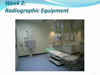
RAD TECH A WEEK 2 RADIOGRAPHIC EQUIPMENT Fall 2010. RADIOGRAPHIC EQUIPMENT. RTA Week 2 Ch. 8 & 9 - pg (110 & 111). Radiographic Room. OBJECTIVES. IDENTIFY GENERIC COMPONENTS OF THE RADIOGRAPHIC EQUIPTMENT DESCRIBE VARIOUS PLANES OF X-RAY TUBE AND TABLE MOVEMENT. THE X-RAY TUBE.

E N D
RAD TECH A WEEK 2 RADIOGRAPHIC EQUIPMENT Fall 2010
RADIOGRAPHIC EQUIPMENT RTA Week 2 Ch. 8 & 9 - pg (110 & 111)
OBJECTIVES • IDENTIFY GENERIC COMPONENTS OF THE RADIOGRAPHIC EQUIPTMENT • DESCRIBE VARIOUS PLANES OF X-RAY TUBE AND TABLE MOVEMENT
THE X-RAY TUBE • The X-Ray tube is the single most important component of the radiographic system. It is the part that produces the X-rays
How “X-rays” are createdSEE: MAN MADE RADIATION (PG.93) TO PRODUCE X-RAYS YOU NEED: • A SOUCE OF ELECTONS • A FORCE TO MOVE THEM QUICKLY • SOMETHING TO STOP THEM SUDDENLY • (More on this week 4)
Radiographic tables • Are designed to support the patient during a radiographic exam • Comfort is not the primary concern • Foam pads should be used if the patient will be required to be on the table for longer than 10 minutes
Tabletop • Must be uniformly radiolucent to easily permit x-ray to pass through. • Carbon fiber is used because it is strong and very little x-ray photons are absorbed. • Usually tabletops are flat however some are curved
Tabletop • Most tabletops are floating, some are motor-driven • The brakes can be released usually by the technologist hand or foot • The brakes are electromagnetic • Floating table tops save significant amounts of time and strain on the technologist
Tables • Tilting rooms are designed for both diagnostic and fluoroscopic work • Tilting models usually tilt to 90 degrees in one direction and 15 – 30 degrees in the other direction • Tilting models include ancillary equipment; footboard, shoulder support, handgrips, compression bands
REMOTE ROOM & OLD CONVENTIONAL FLUORO
Tube Supports • Designed to help technologists with various tube locations for creative imaging. • Tube suspension systems are available in 5 versions: • ceiling mounted, floor-to-ceiling, floor, mobile and c-arm.
Tube Movement • Longitudinal • Transverse • Vertical • Angling or Rolling • Rotating • Telescoping
The ‘BUCKY’ • The bucky is the device in the table or chest board that holds the film cassette. The ‘bucky’ is like a drawer that opens and closes to insert and remove the film cassette.
COLLIMATOR • ATTACHES DIRECTLY BELOW THE X-RAY TUBE • SERVES AS A X-RAY BEAM LIMITING DEVISE • CONTROLS THE SIZE AND SHAPE OF X-RAY FIELD
Field Size • As field size increases, intensity of scatter radiation also increases rapidly. Especially during fluoroscopy
3 Types of beam-restricting devices • Aperture Diaphragm • Cones or Cylinders • Variable aperture collimator
The collimator lamp & mirror • Must be adjusted so that the projected light field coincides with the x-ray beam • Misalignment of the light field and beam can result in collimator cutoff of anatomic structures
ALWAYS KEEP THE COLLIMATED AREA SMALLER THAN THE SIZE OF THE CASSETTE
RADIOGRAPH • PERMANENT RECORD MADE USING RADIATION • RADIO- RADIATION (usually x rays) • GRAPH PERMANENT RECORD (film)
CASSETTE or FILM HOLDER • The CASSETTE is used to hold the film during examinations. It consist of front and back intensifying screens, and has a lead (Pb) backing. The cassette is light tight
CONTROL CONSOLE • GIVES THE TECHNOLOGIST CONTROL OF THE X-RAY MACHINE • TECHNIQUE SELECTION • Located OUTSIDE of the Radiographic Room
The Control Console • The control console is device that allows the technologist to set technical factors (mAs & kVp) and to make an exposure. • Only a legally licensed individual is authorized to energize the console.
“Technique”kVp , mAs (mA x s) • What is set at the control panel • How the “image” is created on the “film” or Image receptor (digital) • kVp controls the “ENERGY” of the beam • The Higher kVp – more penetrating • Ranges is 50 -110 in Diagnostic x-ray
“Technique”kVp , mAs (mA x s) • mA- is the current in combination with the time – determines HOW LONG the beam will stay on • Controls the density on the film/image