Download
1 / 44
510 likes | 1.01k Views
INGUINAL CANAL INGUINAL HERNIA & MALE EXTERNAL GENITALIA. Dr. Mujahid Khan. Inguinal Canal. It is an oblique passage through the lower part of the anterior abdominal wall Present in both sexes It allows structures to pass to and from the testis to the abdomen in males

E N D
INGUINAL CANALINGUINAL HERNIA &MALE EXTERNAL GENITALIA Dr. Mujahid Khan
Inguinal Canal • It is an oblique passage through the lower part of the anterior abdominal wall • Present in both sexes • It allows structures to pass to and from the testis to the abdomen in males • In females it permits the passage of the round ligament of the uterus from the uterus to the labium majus • Transmits ilioinguinal nerve in both sexes
Inguinal Canal • It is about 1 ½ inches or 4cm long in the adults • Extends from the deep inguinal ring downward and medially to the superficial inguinal ring • Lies parallel to and immediately above the inguinal ligament • In the newborn child, the deep ring lies almost directly posterior to the superficial ring
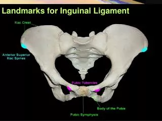
Deep Inguinal Ring • Is an oval opening in the fascia transversalis • Lies about ½ inch (1.3cm) above the inguinal ligament midway between the anterosuperior iliac spine and the symphysis pubis • Margins of the ring give attachment to the internal spermatic fascia
Superficial Inguinal Ring • Is triangular in shape • Lies in the aponeurosis of the external oblique muscle • Lies immediately above and medial to the pubic tubercle • Its margins give attachment to the external spermatic fascia
Anterior Wall of Inguinal Canal • Is formed along its entire length by aponeurosis of the external oblique muscle • It is reinforced in its lateral third by the origin of the internal oblique from the inguinal ligament • This wall is strongest where it lies opposite the weakest part of posterior wall, that is deep inguinal ring
Posterior Wall of Inguinal Canal • Is formed along its entire length by the fascia transversalis • It is reinforced in its medial third by conjoint tendon, the common tendon of insertion of internal oblique and transversus, attached to the pubic crest and pectineal line • This wall is strongest where it lies opposite the weakest part of the anterior wall, that is superficial inguinal ring
Inferior Wall of Inguinal Canal • Is formed by the rolled-under inferior edge of the aponeurosis of the external oblique muscle called inguinal ligament and at its medial end, the lacunar ligament
Superior Wall of Inguinal Canal • Is formed by the arching lowest fibers of the internal oblique and transversus abdominis muscles
Functions of Inguinal Canal • It allows structures of spermatic cord to pass to and from the testis to the abdomen in male • Permits the passage of round ligament of uterus from the uterus to the labium majus in female
Mechanics of Inguinal Canal • The presence of inguinal canal in the lower part of the anterior abdominal wall in both sexes constitutes a potential weakness • Except in the newborn infant, the canal is an oblique passage with the weakest areas, that are superficial and deep inguinal rings
Mechanics of Inguinal Canal • When great straining efforts may be necessary, as in defecation and parturition, the person naturally tends to assume the squatting position • The hip joints are flexed and the anterior surfaces of the thighs are brought up against the anterior abdominal wall • By this means the lower part of the anterior abdominal wall is protected by the thighs
Spermatic Cord • It is a collection of structures that pass through the inguinal canal to and from the testis • It is covered with three concentric layers of fascia derived from the layers of anterior abdominal wall • It begins at the deep inguinal ring lateral to the inferior epigastric artery and ends at the testis
Structures of Spermatic Cord • Vas deferens • Testicular artery and vein • Testicular lymph vessels • Autonomic nerves • Processus vaginalis • Cremastric artery • Artery of the vas deference • Genital branch of genitofemoral nerve
Vas Deferens • It is a cord like structure • Can be palpated between finger and thumb in the upper part of the scrotum • It is a thick walled muscular duct that transport spermatozoa from the epididymis to the urethra
Testicular Artery • It is a branch of abdominal aorta • It is long and slender • Descends on the posterior abdominal wall • It traverses the inguinal canal and supplies the testis and the epididymis
Testicular Veins • These are the extensive venous plexus, the pampiniform plexus • Leaves the posterior border of the testis • As the plexus ascends, it becomes reduced in size so that at about the level of deep inguinal ring, a single testicular vein is formed • Drains into left renal vein on left side and inferior vena cava on right side
Covering of the Spermatic Cord • The covering of the spermatic cord are three concentric layers of fascia derived from the layers of the anterior abdominal wall • Each covering is acquired as the processus vaginalis descends into the scrotum through the layers of the abdominal wall
Covering of the Spermatic Cord • External Spermatic fascia: Is derived from the external oblique aponeurosis and attached to the margins of the superficial inguinal ring • Cremasteric Fascia: Is derived from the internal oblique muscle • Internal Spermatic Fascia: Is derived from the fascia transversalis and attached to the margins of deep inguinal ring
Inguinal Hernia • A hernia is the protrusion of part of the abdominal contents beyond the normal confines of the abdominal wall • Consists of three parts: the sac, contents of the sac, covering of the sac • Hernial coverings are formed from the layers of the abdominal wall through which the hernial sac passes
Indirect Inguinal Hernia • It is the most common form of hernia • Is believed to be congenital in origin • The hernial sac is remains of processus vaginalis • Enters the inguinal canal through the deep inguinal ring lateral to the inferior epigastric vessels • It may extend part of the way along the canal or as far as the superficial inguinal ring
Indirect Inguinal Hernia • If the processus vaginalis has undergone no obliteration, the hernia is complete and extends through the superficial inguinal ring down into the scrotum or labium majus • Under these circumstances the neck of the hernial sac lies at the deep inguinal ring • It is 20 times more common in young males than females • Is more common on the right side
Direct Inguinal Hernia • It composes about 15% of all inguinal hernias • Common in old men with weak abdominal muscles and rare in women • Hernial sac bulges forward through the posterior wall of the inguinal canal medial to the inferior epigastric artery • The neck of the hernial sac is wide
Scrotum • Is an outpouching of the lower part of the anterior abdominal wall • It contains testes, epididymis, and the lower ends of the spermatic cord • Its wall has following layers: skin, superficial fascia, external spermatic fascia derived from external oblique, cremastric fascia derived from internal oblique internal spermatic fascia derived from transversalis, and tunica vaginalis
Skin of the Scrotum • Skin of the scrotum is thin, wrinkled, and pigmented and forms a single pouch • A ridge in the midline indicates the line of fusion of the two lateral labioscrotal swellings • Superficial fascia is continuous with the fatty and membranous layers of the anterior abdominal wall
Superficial Fascia • Superficial fascia is continuous with the fatty and membranous layers of the anterior abdominal wall • The fat is replaced by smooth muscle called dartos muscle • Is responsible for wrinkles of the skin • Innervated by sympathetic nerve fibers • Both layers of sup. Fascia contribute to a median partition that crosses the scrotum and separates the testes from each other
Spermatic Fasciae • Lie beneath the superficial fascia • Derived from three layers of anterior abdominal wall on each side • The external spermatic fascia is derived from external oblique • The cremastric fascia is derived from internal oblique • The internal spermatic fascia is derived from the fascia transversalis
Cremasteric reflex • The cremasteric muscle can be made to contract by stroking the skin on the medial aspect of the thigh, called cremasteric reflex • The function of cremaster muscle is to raise the testis and the scrotum upward for warmth and protection against injury
Tunica Vaginalis • Lies within the spermatic fasciae • Covers the anterior, medial and lateral surfaces of each testis • It is the lower expanded part of the processus vaginalis • Normally shut off just before birth from the upper part of the processus and the peritoneal cavity
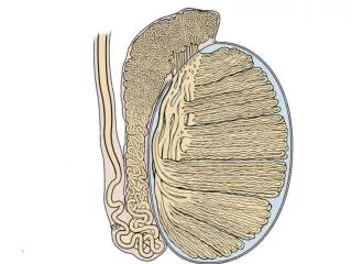
Testis • Is a firm, mobile organ, within the scrotum • Left testis usually lies at a lower level than the right • Upper end of the gland is tilted forward • Surrounded by a tough fibrous capsule, the tunica albuginea • A series of fibrous septa divide the interior of the organ into lobules
Testis • Lying in each lobule are one to three coiled seminiferous tubules • The tubules open into the network of channels called the rete testis • Small efferent ductules connect the rete testis to the upper end of the epididymis
Epididymis • Is a firm structure lying posterior to the testis, with the vas deferens lying on its medial side • Has an expanded upper end, the head, a body, and a pointed tail inferiorly • Lateral groove between it and testis called sinus of epididymis • Is a much coiled tube nearly 20 feet long
Epididymis • The tube emerges from the tail of the epididymis as a vas deferens, that enters the spermatic cord • The long length of the duct of epididymis provides storage space for the spermatozoa and allows them to mature • Its main function is absorption of fluid • May add some substances to the seminal fluid to nourish the maturing sperm