Download

1 / 26
320 likes | 1.19k Views
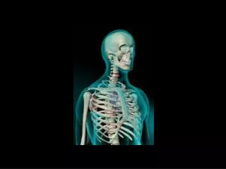
TERMINOLOGY OF THE SKELETAL SYSTEM . I. Regions of the skeleton: a. Cranial b. Postcranial visceral skeleton(=pharyngeal skeleton) vertebral column (including sacrum) ribs sternum b. Appendicular girdles paired appendages unpaired appendages .

E N D
TERMINOLOGY OF THE SKELETAL SYSTEM I. Regions of the skeleton: a. Cranial b. Postcranial visceral skeleton(=pharyngeal skeleton) vertebral column (including sacrum) ribs sternum b. Appendicular girdles paired appendages unpaired appendages
II. Descriptive terms: a. foramen - hole that provides a passage for a vessel or nerve b. canal - tube-like passageway that runs some distance through bone or cartilagec. fissure - cleft-like opening between two bones d. fenestra - opening across which a membrane is stretched or that does not serve primarily as a passageway for a nerve or vessel e. fossa - depression or concavity on the surface of a bone sinus or f. atrium - cavity within a bone, generally of the skull g. process - prolongation or prominence of a part h. condyle - articular prominence i. head - proximal end of a long bone j. apophysis - slender bony protuberance k. tuberosity - large, roughened protuberance usually serving for attachment of a muscle l. fecalcraniated pedagogist - terminology you are undoubtedly applying to the professor of this course by now. You figure out the meaning.
Fibroblasts Meroblasts Scleroblasts Collagen Muscle Osteoblasts Chondroblasts Odontoblasts Ameloblasts Bone Dentin Cartilage Enamel BONE AND MINERALIZED TISSUES I. Origin of Mineralized Tissues Undifferentiated Mesenchyme
II. Mineralized Tissues A. Collagen - precursor to bone and cartilage.Small fibrils bound to form fibersComprise connective tissue in skin and serve as a matrix for formation of bone and cartilage B. Bone 1. Chemical structure Material of collagenous fibers and spaces filled with hydroxyapatite crystals [3Ca5 (PO4)2 . Ca (OH)2]
2. Bone formation a. Membrane and Replacement Bones - • Membrane -form directly from mesenchyme • Replacement or cartilage - formed by replacement of cartilage • Differ only in embryonic origin not in structure Some bones will have both types b. dermal bones - • form from dermal mesenchyme and are homologous with bones that so formed in ancestors • exoskeletal - osteoderms, boney plates of turtles and armadillo • endoskeletal - many bones of skull, pectoral girdle
Typical Bone Formation andStructure Replacement bone
3. Bone architecture Lamellar Bone Typical compact bone - lamellar bone with circular layers of ossified material surrounding a Haversion canal Compact Bone Haversion canal Lacunae Canaliculae
Still Other Mineralized Tissues C. Dentin Similar to bone, but deposited differently - odontoblasts deposit Ca + P not in layers and not in lacunae D. Cartilage - deposition of chondroitin sulfate in a collagenous matrix but no canaliculae and no blood vessels penetrating
2. Types of cartilage a. Hyaline cartilage - abundant in embryos and replaced by bone - ends of long bones b. Fibrocartilage - collagenous bundles intervetebral discs c. Elastic cartilage - elastic fibers - epiglottis, ear cartilage d. Calcified cartilage - Ca salts deposited in skeleton of sharks, especially the jaws e. Mucoid cartilage - gelatinous - rostrum of shark
Other Skeletal Elements D. Tendons - connect muscles with bone E. Ligaments - connect bone with bone F. Sesamoid bones - ossified nodules of bone or cartilage that form in tendons or ligaments
III. Joints - arthroses A. Diarthroses - have synovial sac with synovial fluid • ball and socket - femur • gliding - vertebrae • hinge - elbow • pivot - radius and ulna B. Synarthroses - joints lacking a synovial sac and fluid and are usually immovable 1. synchondroses - junction of cartilage 2. syndesmoses - junction of membrane bone by connective tissue - pubic symphesisAnkylosis - term for fusion of a joint
Neural arch Inter-neural arch Neural canal Pleurocentrum Inter-hemal arch Notochord Inter-centrum Hemal canal Hemal arch THE AXIAL SKELETON Origin - No clear fossil evidence of origin - perhaps because of absence of ossification in adults. I. Basic Components of Vertebral ColumnOne vertebra, probably originally paired - left and right and found in embryos of vertebrates
II. Evolutionary Trends in Vertebral Components of Adults A. Loss of intercalary arches 1. Most vertebrates have only neural arch with hemal arch present only in caudal vertebrae2. Interneural and interhemal arches lost in all tetrapods but present in cyclostomes, chondricthyians, dipnoi (lungfish) and chondrososteiLoss of intercalary arches provided increased flexibility
B. Loss of hemal arches occur in fish and amphibians, well developed in reptilespoorly developed in birds and mammals primarily caudal vertebrae and remain as chevron bones
C. Reduction of number of centra 1. Types of reduction - reduces to one = monospondyly or none = aspondyly dispondyly remains in some fish Shark trunk vertebrae
2.Modes of reduction a. fusion of pleurocentra and intercentra (common in fishes) b. incorporation of intercentra into intervertebral bodies (trunk vertebrae of amniotes except atlas and axis) c. transformation of the intercentrum into parts of chevron bone in tail of amniotes d. others too
Anterior D. Formation and fate of inter vetebral bodyOriginally, vertebrae form around notochord, centrum encircling it and arches rest on top.Perforate amphicoelous - fish and amphibians except frogs.In all others, notocord restricted to intervertebral bodies. Fate of these variesImperforate amphicoelous - some birds, reptiles and non-frog amphibiansOpisthocoelous - some reptilesProcoelous - snakes, lizards, and most birdsAcoelous - mammals Perforate Amphicoelous Imperforate Amphicoelous Notochord Procoelous Opisthocoelous Acoelous
Boney Fish - Amphicoelous Snake - Procoelous Bird - Procoelous (Heterocoelous) Mammal - Acoelous
III. Regionalizationof the Vertebral Column A. Earliest Vertebrates - trunk and caudal typical of fishes B. Early tetrapods (cervical and sacral first appear in amphibians - Permits more cranial mobility No ribs C. Higher tetrapods (cervical, sacral, and thoracic and lumbar in reptiles, birds, mammals)
IV. Neck Vertebrae Atlas - no centrum - with skull attached to the axis as odontoid process.
V. Sacrum and Synsacrum support pelvic girdle and are fused sacral vertebraeSynsacrum - In birds, sacrum plus first few caudals and fused to ilia of pelvic girdle
VI. Caudal Vertebrae - several in most vertebrates fused in birds - pygostyle coccyx - apes
VII. Ribs A. Fish - Dorsal and ventral in some fish - salmon ventral in most fish B. Tetrapods - primitive forms have bicepital ribs Capitum - antero-ventral articulates with parapophysis Tuberculum - postero-dorsal articulates with diapophysis on the neural arch Uncinate processes in birds - connection between two ribs
VIII. Sternum • In primitive tetrapods, served for protectionsegmented and later became a solid structure • Ribs were originally free, only later do they articulate with sternum • Some articulate with pectoral girdle • Form major part of turtle shell