Download
1 / 35
350 likes | 1.01k Views
Use of a Radiograph to Assist with Therapeutic Shoeing and Trimming. Debra R. Taylor, DVM, MS, DACVIM Associate Professor Department of Clinical Sciences College of Veterinary Medicine Auburn University. Equine Podiatry. The specialty dealing with the equine hoof the hoof capsule

E N D
Use of a Radiograph to Assist with Therapeutic Shoeing and Trimming Debra R. Taylor, DVM, MS, DACVIM Associate Professor Department of Clinical Sciences College of Veterinary Medicine Auburn University
Equine Podiatry • The specialty dealing with the equine hoof • the hoof capsule • coronary band • all anatomical structures with in the hoof capsule • all tendons that affect the hoof • Requires the veterinarian and trimmer or farrier to work together
What does the horse need? • A veterinarian and a trimmer or farrier team who are willing to work together • A team that enjoys podiatry and is committed to doing their best work
What does a farrier or trimmer need from the veterinarian? • A veterinarian • who understands what is healthy and what is not • who understands diseases affecting the hoof • who can consistently take an informative radiograph of the foot • who is willing to work as part of a team • Take the time to show him the radiographs • Pre- and post- trimming or shoeing • Learn to speak the same language • Many overlapping terms
Why do you need a radiograph to monitor shoeing or trimming a horse? “Quality radiographs are to podiatry what ultrasound is to reproduction” Learning curve Without ultrasound or radiographs With ultrasound or radiographs Ability to make a positive change Time Time
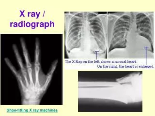
Traditional Radiography of the digit Goals • To evaluate the proximal and distal interphalangeal joints, integrity of bony structures, and to view radiographic degenerative changes in the foot.
Traditional Radiography • Beam focused at coronary band • Foot packed • Ground surface not always demarcated • Dorsal hoof wall marked with rigid object • Apex of frog and heels not marked • Hoof not always directly against cassette
Radiographic GOALS • Consistent informative films • Take the same view every time • Ability to monitor small changes with accuracy • Evaluate the soft tissues of hoof • Evaluate P3’s relationship to capsule and ground • Evaluate how the bone and the soft tissues relate to each other • How the bone and soft tissue relate to the ground • Learn common parameters of breeds • What is common is not necessarily healthy • Horses have only rarely been bred for hoof quality • Popular breeds are notorious for poor hoof health
Radiography for Shoeing and Trimming • Center on bottom of P3, not coronary band • measure sole depth • Palmar/plantar angle • medial to lateral balance of P3 • Clearly demarcate (with barium paste) • Dorsal hoof wall – care to start paste exactly on hairline • Lateral heel bulb – care to start paste exactly on hairline • Apex of frog • Clearly demarcate ground surface – lead in wooden block • Hoof touching cassette • Heel bulbs parallel to primary beam and perpendicular to cassette • Do not pack the foot • Measure sole depth • Standardize distance from film to machine
Methods • Center on bottom of P3 • Blocks match machine • metal wire in top • Barium paste on dorsal hoof wall • Barium on apex of frog • Barium on heel
Much better • Centered bottom of P3 • Dorsal hoof wall marked • Ground Surface Marked • Not packed • Heel marked • Apex marked • Note: -ok that bone is too light, not as worried about boney detail here as relationship of the bone to the soft tissues and ground NOTE: Trimmers and farriers appreciate viewing the hoof in the direction it would be if they were looking at the horse, RF point toe to right, LF point toe to left
Equine Podiatry Radiology Primary beam is 1 cm off top of blocks Build blocks to match your machine so can Set the machine on the ground and horse Will be in correct positions NOTE: need two blocks So horse is standing level
Breakover Breakover
Podiatry Radiology Measurements • Breakover • - The horizontal distance from the tip P3 to where the hoof “breaks over” or pivots. • 0 – 3.5 cm is ideal, depending on the horse • optimizing breakover • decreases sheer force on P3 and hoof wall • decreases tendon and muscle strain
Podiatry Radiology Measurements • Palmar Angle • The angle that is formed from the palmar/plantar surface of P3 and the ground. - 2- 10° degrees normal (Adams)
Podiatry Radiology Measurements • Sole Depth • Soft tissue opacity just distal to the plantar surface of P3 • The sole should be greater than 10mm in depth.
Podiatry Radiology Measurements • Horn: Laminar Zone • Distance from the dorsal hoofwall to the dorsal surface of P3. • Measured at both the dorsal proximal and dorsal distal ends of the foot. • Should be uniform and < 20 mm
Podiatry Radiology Measurements • CE Zone • - The CE Zone is the vertical distance between the Coronary Band and the Extensor Process. • Ideally < 1cm. • Need great care in marking the hairline, because plan to monitor change in this number
Podiatry Radiology Measurements • Cup • The radiolucent area distal to the sole • Measure to the ground surface • Some horses have none
Podiatry Radiology Measurements • Bone Angle • Angle formed at the junction of the dorsal and palmar/plantar surface of P3
Evaluation of Lateral View • Horn: Laminar Zone • Should be uniform ( < 20 mm) • CE zone - <1 cm • coronary band - extensor process • “halo effect”- coronary band invagination • Sole depth • mass (> 10 mm) • Cup • Black area between sole and ground surface
Evaluation Lateral View • Palmar Angle • Angle formed between palmar surface of P3 and ground • The horse wants it positive 3-9 º • Digital Breakover • distance from the tip of P3 to where hoof breaks forward • The horse wants it short, maybe even negative? • The horse wants it optimized. • Bone Angle • angle formed between dorsal and palmar surfaces of P3
Anterior – Posterior projection • Looking for medial lateral balance of P3 • Uniformity of joint spaces • Is the medial and lateral coronary band level • Evaluate growth rings if use barium
Moral • TAKE the radiograph the correct way • Evaluate the parameters • Use the film to make goals • Evaluate whether goals were met • Work together!!!!