Download
1 / 42
420 likes | 762 Views
Ch. 16 DNA: The Genetic Material Intro In 1953, James Watson and Francis Crick presented their model of DNA to the world. Nucleic Acids (DNA: Deoxyribonucleic acid, RNA: Ribonucleic acid) have a unique ability to replicate itself. DNA’s ability to replicate itself precisely is

E N D
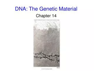
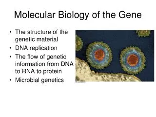
Ch. 16 DNA: The Genetic Material
Intro • In 1953, James Watson and Francis Crick • presented their model of DNA to the world. • Nucleic Acids (DNA: Deoxyribonucleic acid, • RNA: Ribonucleic acid) have a unique ability • to replicate itself. • DNA’s ability to replicate itself precisely is important for its transmission from one generation to the next. • The search for genetic material led to the • discovery of DNA and its structure. • Before the 1940’s it was thought that • proteins were the genetic material.
The genetic role of DNA was first researched • by Frederick Griffith in 1928. • Studied Streptococcuspneumoniae, a • bacterium that causes pneumonia in • mammals. • He discovered one strain that was • nonvirulent (harmless) - R strain. • Another strain was virulent (causes • pneumonia) – S strain. • His experiment: • Mixed heat-killed S strain with • live R strain and injected it into mice.
The mouse died and Griffith took a • blood sample. • He found that some of the R strain • had changed into the S strain. He • called this transformation. Some • chemical component had changed the • R strain into S strain.
After Griffith’s experiment, researchers tried to discover this transforming material. • Finally in 1944, Oswald Avery, Maclyn • McCarty and Colin MacLeod announced that • the transforming substance was DNA. • They took various chemicals from the • heat-killed pathogenic bacteria and tried • to transform nonharmless bacteria with • them. Only DNA worked. • In 1952, Alfred Hershey and Martha Chase • showed that DNA was the genetic material • of the phage T2 (a virus that infects • e. coli bacteria).
They studied bacteriophages – viruses • that infect bacteria. They knew that • viruses need bacteria in order to • replicate. • Since viruses have simple structure, they • wanted to know whether it was their • protein coat or DNA that was the genetic • material.
Their experiment: • They had two batches of viruses: • -Viruses with radioactive sulfur (S-35) • labeling their protein coat. • -Viruses with radioactive phosphorus • (P-32) labeling their DNA. • They allowed for the two batches to • infect bacteria. After infection, they • put the virus/bacteria mixture in a • blender so that the viral parts outside • of the bacteria could be separated.
They centrifuged the mixture so that • the bacteria form a pellet at the • bottom of the tube. • Then they tested the bacteria for • radioactivity. • Found radioactivity inside bacteria.
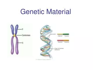
They concluded that the injected DNA, • radioactively labeled was the genetic • material. • In 1947, Erwin Chargaff had developed a • series of rules based on a survey of DNA • composition in organisms. • He already knew that DNA was a polymer • of nucleotides consisting of a nitrogenous • base, deoxyribose, and a phosphate • group. • The bases could be adenine (A), thymine • (T), guanine (G), or cytosine (C). • Chargaff noticed that the DNA • composition varied from species to • species.
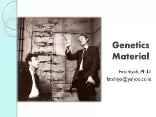
He found that the bases were present in • all species in very regular ratios: -The number of Adenine = Thymine -The number of Cytosine = Guanine • Watson and Crick: By the 1950’s it was now • accepted that DNA was the genetic material. • The race was on to discover its structure. -Linus Pauling -Maurice Wilkins and Rosalind Franklin
Wilkins and Franklin used X-Ray crystallo- • graphy to study the structure of DNA. • From their picture • of DNA, Watson • and Crick were able • to see its helical • structure. • Double-Helix model of DNA proposed by • Watson and Crick: • DNA is made up of nucleotides.
2. DNA looks like a ladder. It has two strands, each strand with the sugar- phosphate chains on the outside and the nitrogenous bases on the inside. • The nitrogenous bases paired up, forming • the rungs of the ladder. • The ladder is then twisted, forming a coil.
The nitrogenous bases are paired up very • specifically: • A – T • G - C pyrimidines (single ring) purines (double ring) -Only a pyrimidine-purine pairing would produce the 2-nm diameter indicated by the X-ray data.
-The A & T, C & G form hydrogen bonds between one another: -A = T (two) -G = C (three) ** This confirms Chargaff’s observations.
The Structure of DNA • http://www.sumanasinc.com/webcontent/anisamples/molecularbiology/DNA_structure.html
The sequence of nucleotides on one DNA • strand can vary in numerous ways. Each • gene has a specific sequence of • nucleotides. A portion of gene has the following sequence of nucleotides: A T G G A C T T C • T • A • C • C • T • G • A • A • G -Watson and Crick presented their DNA model in 1953. -They, along with Maurice Wilkins won the Noble Prize in Medicine in 1962.
Crick Watson
DNA Replication: • After Watson and Crick presented their DNA • model, they wrote about how DNA replicates. • They said that the two strands of DNA • are complimentary to one another. • When they are separated, they can act • as templates for synthesizing a new • strand of DNA.
Watson and Crick’s model of replication • was called “Semiconservative replication.” -This means that when two strands of DNA are made, each one will have a new strand and an old one. The old strands will act as “templates” to the new complimentary strand. • Experiments done in the late 1950s by • Matthew Meselson and Franklin Stahl • supported the semiconservative model. 1. In their experiments, they labeled the nucleotides of the old strands with a heavy isotope of nitrogen (15N) while any new nucleotides would be indicated by a lighter isotope (14N).
After they labeled the DNA and let it • replicate, they found that each DNA • molecule had one strand labeled with • N-15 and the other with N-14. • They then allowed for the DNA to • replicate once more and they found that • the only strands with N-15 were the • original two strands of DNA.
Meselson-Stahl Experiment • DNA Replication • http://www.sumanasinc.com/webcontent/anisamples/majorsbiology/meselson.html
More than a dozen enzymes and proteins • carry out DNA replication: • E. coli can replicate its DNA in less than • an hour. • Human cells can replicate its 6 billion • base pairs in only a few hours. • Replication is highly accurate; only one error per billion nucleotides. • DNA replication starts at the origins of • replication. • In bacteria, there are very specific • nucleotide sequences that enzymes • recognize as sites where replication • begins.
2. In eukaryotes, there are many sites on the DNA strand where replication takes place. • At the origin of replication, a replication • bubble forms, where new DNA strands • are elongated in both directions.
DNA Polymerase is the enzyme that • elongates the new DNA at a replication • fork. • The rate of elongation is about 500 • nucleotides per second in bacteria and • 50 per second in human cells. -The nucleotides that are attached to the newly formed strands are called nucleoside triphosphates. Each has a nitrogen base, deoxyribose, and a triphosphate tail.
-As each nucleotide is added, the last two phosphate groups are hydrolyzed to form pyrophosphate.
-The exergonic hydrolysis of pyrophosphate to two inorganic phosphate molecules drives the polymerization of the nucleotide to the new strand.
The strands in the double helix are • antiparallel. • The sugar-phosphate • backbones run in • opposite directions. • One strand goes • from 3’ 5’ • direction. The • other strand goes • from 5’ 3’ • direction.
DNA polymerases can only add • nucleotides to the free 3’ end of a • growing DNA strand. -DNA can only replicate in the 5’ 3’ direction.
-Leading strand replicates from 5’ 3’. -Lagging strand replicates from 5’ 3’, but by forming Okazaki fragments. -The Okazaki fragments (100-200 nucleotides), are then joined by DNA ligase.
DNA replication starts with a primer (a • short fragment of RNA). • A primer is • created by an • enzyme called • primase. • Once the primer • is made, DNA • polymerase can • start adding • nucleotides at the • 3’ end. • The primer is • then converted • into deoxyribo- • nucleotides.
Only one primer is • needed for the • leading strand. • A new primer is • needed for each • Okazaki fragment.
Enzymes involved in DNA replication: • 1. Primase: • 2. DNA Polymerase: • 3. DNA Ligase: Creates a primer. Adds nucleotides to the 3’ end; replaces RNA primer. Joins the Okazaki fragments. • Helicase: Untwists DNA and separates • the template strands at the replication • fork. • Single-strand binding proteins: keep • the template strands apart during • replication.
Enzymes proofread DNA during replication • and repair existing damaged DNA. • Mistakes during DNA synthesis can • occur at a rate of one error per 10,000 • base pairs. • DNA Polymerase proofreads the new • DNA strand. If there is a mistake, DNA • polymerase removes the incorrect • nucleotide and resumes synthesis. • After proofreading, the error rate is • one per billion nucleotides.
Harmful chemicals, radioactive emissions, • X-rays, and ultraviolet light can change • nucleotides. Also, under normal cellular conditions, DNA can undergo spontaneous mutations. • There are over 130 enzymes that help • repair damaged and mutated DNA. • Defects in enzymes that help repair • mismatched nucleotides are associated • with colon cancer. • Nucleases are enzymes that excise • (cut out) damaged nucleotides. After • they are cut out, the gap is filled in • with the correct nucleotide via DNA • polymerase and ligase.
Example: The inherited disorder called Xeroderma Pigmentosum causes an individual to be very sensitive to sunlight. UV light can cause two adjacent Thymine nucleotides to form a dimer. The dimer buckles the DNA strand and interferes with DNA replication. Causes skin cancer.
The End-Replication problem: When • eukaryotic DNA replicates, a gap is left at • the 5’ end of each new strand because DNA • polymerase can only add nucleotides at the • 3’ end. Gap formed where primer previously existed.
To help this problem, eukaryotic DNA • have telomeres at their ends. Telomeres • are not genes but the sequence • TTAGGG • repeated between 100 to 1,000 times. • The telomeres prevent any important • genes from being deleted over time due • DNA shortening with repeated replication. • The enzyme, telomerase, catalyzes the • lengthening of telomeres.
-Telomerases have a short RNA fragment that serves as a template for a new telomere.