Download

1 / 61
610 likes | 978 Views
Arm Positioning and Screw Placement in Massive Rotator Cuff Tears. WILLIAM F BENNETT MD Sarasota, Florida. Purpose.

E N D
Arm Positioning and Screw Placement in Massive Rotator Cuff Tears WILLIAM F BENNETT MD Sarasota, Florida
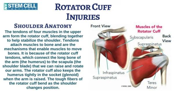
Purpose • Illustrate Arthroscopic Techniques to Facilitate- a) identification of retracted tendons b) separation of tendon from bursae c) separation of tendon from glenoid d) mobilization of retracted tendons e) arm positioning and mobilization f) arm positioning and screw placement g) arm positioning and suture placement h) arm positioning and knot tying Keep in mind that chronic retraction and fatty degeneration may indicate a situation in which the cuff is not repairable
a) Identification of retracted tendons • Prior to repair it is important to identify all structures torn • initial visualization of tendons from glenohumeral view can help plan the repair • repair of all tendon tears without repair of coracohumeral head tears may still result in unwanted medial lateral motion of the biceps tendon
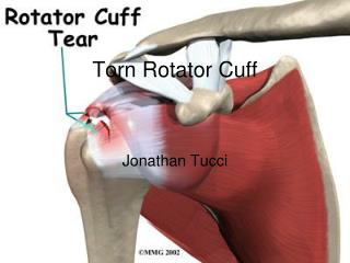
a) Identification of retracted tendons(contd) • In large and massive tears crescent appearance typical from glenohumeral and subacromial view • Individual tendons vary from mainly subscapularis and supraspinatus to supraspinatus and infraspinatus to subscapularis, supraspinatus and infraspinatus • Degree of retraction varies • Involvement of heads of the coracohumeral ligament vary View from lateral portal
a) Identification of retracted tendons cont’d • Infraspinatus retracts not only medially but postero-inferiorly, best visualized from subacromial space • posterior to spine of scapula • usually is scarred into the inner fascia of the deltoid muscle • very important plane between infraspinatus and posterior deltoid • often difficult to differentiate tendon from bursae from inner fascia of deltoid
a) Identification of retracted tendons cont’d • Supraspinatus often retracted to glenoid level • can be visualized from glenohumeral and subacromial space • anterior to spine of scapula • recognize that while a large crescent sign is present, there is usually a component of longitudinal splitting between supraspinatus and infraspinatus • while longitudinal split may not be visible often side-to-side repair is needed to achieve coverage
a) Identification of retracted tendons cont’d • Subscapularis tendon can vary in its involvement from partial thickness and length to full thickness and length • best visualized from glenohumeral joint with arm in flexion and internal rotation • the lateral head of the coracohumeral ligament must be disrupted to have retraction of the subscapularis tendon • IASS may be disrupted yet subscapularis appears in relative anatomic position • the subscapularis tendon is involved approximately 30% in all rotator cuff tears to some degree
* IASS may be disrupted yet subscapularis appears in relative anatomic position * The subscapularis tendon is involved approximately 30% in all rotator cuff tears to some degree * MRI findings are subtle but present with subscapularis tears a) Identification of retracted tendons cont’d
a) Identification of retracted tendons cont’d • Biceps subluxation can occur with varying combinations of rotator cuff tendon involvement • Supraspinatus and lateral head coracohumeral ligament • Subscapularis and medial head coracohumeral ligament • Supraspinatus , subscapularis and both heads of the coracohumeral ligament constitutes complete disruption of the bicipital sheath • Thus, important to identify structures from the glenohumeral view
b) Separation of tendon from bursae • Posteriorly- must take judicious time to separate infraspinatus from inner fascia of posterior deltoid • technique requires placing a shaver with closed end against the tendon and under direct arthroscopic visualization sweeping the shaver downwards while applying pressure against the infraspinatus • in time there will be a space identified between the infraspinatus tendon and deltoid
b) Separation of tendon from bursae cont’d • Final visualization will allow one to see the insertion of the teres minor and visualize the muscle tendon junction of the infraspinatus • The separation must be taken inferiorly to a sufficient level to mobilize the entire infraspinatus as it usually is balled up and subluxed postero-inferiorly
b) Separation of tendon from bursae cont’d • Superiorly the supraspinatus tendon is retracted and typically retracted to the glenoid level and anterior to the spine • It is contiguous with the coracohumeral ligament and separation of the tendon from overlying bursae and fat pads will help identify both • separation of the tendon from the fat pad is essential
b) Separation of tendon from bursae cont’d • Anteriorly the subscapularis may or may not be retracted • Repair of partial and full thickness partial and full length tears without retraction typically require a portion of the body of the coracohumeral ligament to be resected for visualization, area of the anterior portal • Retracted tears require the same shaver technique to remove the subscapularis from the inner fascia of the anterior deltoid
c) Separation of tendon from glenoid • Posteriorly- the infraspinatus can be separated from the glenoid from the lateral portal with visualization from either anterior or posterior • keep in mind that the suprascapular nerve is not far away • Superiorly-hardest to mobilize any length from anterior glenoid • Anteriorly-lateral portal, plane between the coracohumeral ligament and coracohumeral ligament
d) Mobilization of retracted tendons • Mobilization should be maximized so as not to place the tendons under too much tension • mobilization is more than separation • often either a stay suture of a soft tissue grasper helps to mobilize the tendons to the footprint of the rotator cuff insertion • mobilization requires that all tendons be free from glenoid and from overlying structures
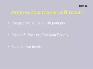
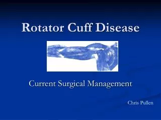
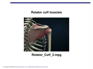
Posterior view Lateral view
E) Arm positioning in conjunction with mobilization • Beach Chair position allows for many degrees of freedom during the repair • the use of a Mayo stand(elevation) aids in bringing the arm into abduction in order to bring the arm to the tendons rather than the tendons to the arm • With the arm at 60-80 degrees of abduction the arm can be internally and externally rotated to bring the various portions of the rotator footprint to approximation with the tendons
E) Arm positioning in conjunction with mobilization cont’d • Abduction and external rotation facilitates approximation of infraspinatus with footprint • further abduction facilitates approximation of the supraspinatus tendon with footprint • abduction and internal rotation facilitates approximation of the sleeve of the coracohumeral ligament
E) Arm positioning in conjunction with mobilization cont’d • Subscapularis mobilization and repair is best done from the glenohumeral joint • Often the IASS and/or MCHL is torn • fibers often remain attached traversing to lateral bicipital sheath • shoulder flexion and internal rotation helps visualize these lesions • a soft tissue grasper through the subacromial lateral portal can be placed through the supraspinatus defect and this tissue reduced to proper footprint
E) Arm positioning in conjunction with mobilization cont’d • Preparation of bed of bleeding bone for IASS attachment(subscapularis) best done through superior portal with arm in flexion and internal rotation • arm position for attachment should be with the arm at side and arm held at external rotation which is equal to opposite side
F) Arm Positioning and Screw Placement-infraspinatus and supraspinatus • Screw should be inserted at 45 degrees to bone • arm should be closer to side to facilitate proper angle • use one 5mm corkscrew with #2 tevdek by 2 for each centimeter of tear • start from either far anterior or far posterior and work away from first screw • I use a portal directly through the skin with no cannula placed directly anterior to the lateral subacromial portal • I place all screws through same hole • Vary screw placement by movement of arm in internal and external rotation
F) Arm Positioning and Screw Placement-infraspinatus and supraspinatus cont’d • Posteriorly- first screw should be at insertion of teres minor • best visualization of this insertion with arm abducted 60-80 degrees and in full internal rotation • However, screw placement may require that the arm be brought out of abduction to achieve “dead-man” angle
F) Arm Positioning and Screw Placement-infraspinatus and supraspinatus cont’d • Place all screws at once • through same skin incision • proceed anterior from posterior to anterior by externally rotating the arm in 60-80 degrees of abduction • often trial reduction of all tendons is necessary with soft tissue grasper to visualize reduction
F) Arm Positioning and Screw Placement-subscapularis • Subscapularis screw placement is different • Screws are placed through the anterior portal with the scope in the glenohumeral joint • If retracted screws go directly into the previously prepared bed of bleeding bone • if not retracted screws go through the tendon after noting insertion site with trial reduction • screw is brought through tendon and into the joint under direct visualization and then backed out • then the arm is brought into proper external rotation and the insertion device is used to lever the subscapularis into position and the screw is advanced into the bone
g) Arm positioning and suture placement • Sutures nonabsorbable #2 Tevdek preferred • tendon edges debrided to viable tissue • passage with shuttle relay or disposable suture retriever • introduced through lateral portal • retrieved through anterior portal • then pass through limb and post brought through lateral portal • various combinations of Mayo stand elevation and internal and external rotation allow for proper placement of sutures through tendons
h) Arm positioning and knot tying • Various combinations of Mayo stand elevation and internal and external rotation of the shoulder allow for knots to be tied by bringing bone to tendon rather than having to bring tendon to footprint and hold in place • alternative is to mobilize the tendon to footprint and hold in place with soft tissue grasper or by passing a stay suture first
h) Arm positioning and knot tyingcont’d • First knot should secure the teres minor infraspinatus junction • usually use two knots in close proximity • knot is tied with arm abducted 60-80 degrees and shoulder brought into external rotation which brings the footprint to the posterior tendons • proceed from posterior to anterior by incrementally bringing the shoulder from external rotation to internal rotation-see next slide • sometimes it is easier to secure anterior and posterior margins of the “crescent” then work on the middle portion of the tear-see 2nd following slide
h) Arm positioning and knot tyingcont’d • Side-to-side repair is facilitated by passing a free #2 Tevdek from the anterior portal through the anterior tendons across the longitudinal split and out through the infraspinatus tendon • space developed between the inner fascia of the posterior deltoid and the infraspinatus tendon will allow for the suture to be directly visualized and retrieved through the lateral portal
h) Arm positioning and knot tyingsubscapularis • Knots are retrieved through the superior portal and then brought out through anterior cannula • dual monitor helps facilitate tying by allowing the surgeon to come to the anterior part of the shoulder and tie directed posterior • remember in this position the monitor image is a mirror image and the anatomic area you are visualizing or tying will require you to move you hand 180 degrees to the opposite side of what you think