Download
1 / 57
610 likes | 2.01k Views
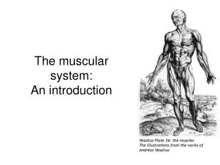
The muscular system: An introduction . What is it?. Collection of ~600 skeletal muscles Also many smooth muscles and heart tissue Myology: study of muscle Myo-, mys-, sarco- all refer to muscle. What do muscles do for me?. Move you and your internal organs Provide stability

E N D
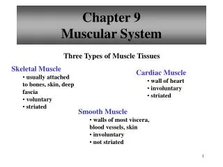
What is it? • Collection of ~600 skeletal muscles • Also many smooth muscles and heart tissue • Myology: study of muscle • Myo-, mys-, sarco- all refer to muscle
What do muscles do for me? • Move you and your internal organs • Provide stability • Maintain posture • Control body openings/passages • Produce body heat (~85% of it)
What are the parts of a muscle? • Watch animation on your CD • Muscle cell = muscle fiber • long (up to 30 cm) • Epimysium: • Perimysium and fascicles • Fascicle = group of muscle fibers • Deep vs. superficial • Endomysium • CT sheaths = for blood vessels and nerves
What are the characteristics of muscle tissue? • Excitability: ability to receive/respond to stimuli • Receive neurotransmitter (e.g. acetylcholine) • Response: contract • Contractility: ability to shorten • Extensibility: ability to be stretched/extended • Elasticity: ability to recoil
Where do muscles attach to bone? • Attached in a minimum of two places • Insertion: point which moves toward immovable bone • Origin: point of insertion at immovable bone • Limbs: usually origin is proximal to insertion • Also it (or its tendon) MUST cross a joint between the origin and insertion! • If a muscle (or its tendon) didn’t, what would happen?
How do muscles attach to bone? • Direct (fleshy) attachment • epimysium fused to periosteum or perichondrium
How do muscles attach to bone? • Indirect attachment • Collagen fibers of epimysium become tendon • Tendon merges with periosteum • Much more common • Aponeurosis: sheetlike tendon connection • Scalp, abdomen, lumbar, hand, foot
How do muscles move together? • Agonist(prime mover) • Synergist helps • Antagonist: Opposes prime mover • Antagonistic pair:act on opposite side of joint • Fixator: e.g. to scapula so biceps move radius and not scapula • muscle movement movie
What special features are found in muscle fibers? • Multiple nuclei: fusion of many myoblasts • Satellite cells nearby • Sarcolemma • Sarcoplasm • Many glycosomes (with glycogen) • Lots of myoglobin • Stores oxygen like hemoglobin
What special features are found in muscle fibers? • Sarcoplasmic reticulum (SR):releases calcium • Myofibrils: many in each myocyte • long, contractile elements, parallel to myocyte length • Contains many myofilaments
How do muscles contract? • First, look at anatomy of myofilaments • Appears as a striation pattern • Sliding filament animation
What creates the striations? • Alternating A (dark) and I (light) bands • A band • H zone: light zone in middle • M line • Only visible when muscle relaxed • I band • Z disc: dark line in middle • Sarcomere = distance between two Z disks
What three myofilaments are found in myofibrils? • Thick filaments: myosin • Extends the length of A band • Looks like golf club with two heads (cross bridges)
What three myofilaments are found in myofibrils? • Thin filaments: actin • Extend across I band, partially into A band • Anchored to Z disk • G actin subunit is binding site for myosin head • Tropomysin lines actin grooves • Troponin bound to tropomysin; binds calcium
What three myofilaments are found in myofibrils? • Elastic filaments: titin (AKA connectin) • Strong recoil, found in center of thick filament • Anchor thick filament to Z disc
What do all these proteins do? • Contractile proteins • Myosin and actin • Workhorses • Regulatory proteins • Tropomysin and troponin • Control when contraction happens • Based on calcium availability
What determines calcium availability? • Sarcoplasmic reticulum • Smooth ER, forming parallel tubules • Tubules surround each myofibril • Stores calcium, releases it when NERVE demands it • Terminial cisternae: perpendicular ER tubules
What determines calcium availability? • T tubules: at A band-I band junction • Extension of sarcolemma • Extends as tubule through paired terminal cisternae • Forms triad • Transmits nerve signal deep into myocyte • Stimulates calcium release from sarcoplasmic ret.
So how do all these parts make muscles contract? • Sliding filament theory • Time for that CD! • Result: thin filaments slide along thick filaments causing contraction
How do nerves “tell” muscles to contract? • Via action potentials • Changes in electrical charge across sarcoplasm • Stimulates calcium release • action potential and contraction animation • Called Excitation-Contraction Coupling • First, we need to know more about neuromuscular junctions and action potentials
Where do nerve and muscle touch? • Neuromuscular junction • Motor neuron stimulates skeletal muscle • Axon divides to form numerous neuromuscular junctions • One neuromuscular junction per muscle fiber • Motor unit = all myocytes innervated by one nerve (20-1,000) • More motor units = finer control • Muscles have > 1 motor unit to prevent fatigue
Where do nerve and muscle touch? • Synaptic cleft separates muscle fiber from nerve • 60-100 nm space • Synaptic knob (end of axon) • Motor end plate (depression in muscle fiber)
If nerve and muscle don’t touch, how do they communicate? • Nerve impulse reaches axon • Voltage-regulated calcium gates open, cause • Synaptic vesicles to be exocytosized • Release acetylcholine (ACh) • ACh traverses synaptic cleft • ACh receptors on myocyte bind Ach • Stimulates opening of calcium gates • Action potential propagates down T tubules • Calcium released to stimulate contraction
What is a polarized cell? • All cells are polarized (resting potential) • Differential charge across PM • High K+ inside, high Na+ outside • Also DNA, RNA high negative charge • Overall, inside of cell is neg., outside pos. • resting potential animation
What are action potentials? 1. Depolarization • When ACh binds to ACh receptors, Na+ sensitive gates open • Inflow of Na+ changes charge (voltage) difference across membrane
What are action potentials? 2. Propagation • action potential spreads across sarcolemma • Opens voltage-sensitive Ca2+ gates
What are action potentials? 3. Repolarization • K+ gates open to reestablish charge • Refractory period: time its takes to reestablish charge • Action potential movie
How does a cell return to its resting state? • After several rounds of depolarization, too much Na+ on inside • Na+/K+ pumps • Export 3 Na+ and import 2 K+
Meanwhile, back at the synaptic cleft… • Acetylcholinesterase (AChE) destroys • any ACh still in synaptic cleft • Prevents continual stimulation
TO REVIEW:What happens during excitation-contraction coupling? Excitation • Action potential travels along sarcolemma and down T tubules • Action potential reaches triad • causes terminal cisternae to release calcium into sarcoplasm • Calcium bind to troponin, moves tropomyosin out of the way
What happens during excitation-contraction coupling? • Contraction • Myosin heads attach to actin and pull thin filaments toward H line • Attachment, power stroke, reattachment, cocking • Relaxation • Within 30 ms, calcium removed • via ATP-driven calcium pump • At same time AChE degrades ACh • Tropomyosin blockage reestablished • cross bridge activity ceases
What makes a contraction strong? • Length-tension relationship • Too little = ___________ contraction • Too much overlap = contraction ___________ • Optimal overlap allows for greatest contraction • CNS maintains constant, partial contraction: tonus (muscle tone)
How do whole muscles work? • Threshold: minimum voltage for contraction • Stimulate a nerve or a myocyte: • Latent-period: delay before contraction (for excitation, etc.) • Higher voltage does not produce stronger contraction
So how do muscles contract at varying strengths? • Recruitment • Multiple motor unit summation • Temporal summation • Produces treppe (staircase phenomenon) • If enough stimuli fast enough, produces incomplete tetanus • Why? • Stimuli too rapid to clear calcium between contractions? • Heat released causes enzymes to work more efficiently (e.g. warm-up exercises)?
What happens if muscle stimulated too much? • Individual contractions fuse to smooth contraction • Complete tetanus • Different than the disease tetanus • This blocks glycine (an inhibitor) release
Where do muscles get the ATP needed to contract? • Anaerobic fermentation • Pro: don’t need oxygen to make ATP • Cons: only makes a little ATP and produces lactic acid • Aerobic respiration • Pro: much greater ATP yield • Con: requires oxygen
How much ATP do muscles need? • Yes, if you only want to shorten your muscle by about 1% • Cycle repeated until desired shortening reached • Usually this is about 30 to 35% • Note: always some myosin heads attached • Prevents thin filament from sliding back • Cycle stops if SR pumps calcium back out of sarcoplasm • Also stops if no more ATP
What if there’s no ATP? • Rigor mortis • Starts at 3 to 4 hrs after death • Peaks at 12 hours • Dissipates over next 48 to 60 hrs • Calcium no longer pumped out • Myosin stays stuck to actin • ATP runs out shortly after person stops breathing • No ATP means myosin heads can’t detach • Rigor mortis disappears as muscle proteins break down • Related to liver mortis
When do muscles use each ATP-generating path? • Immediate demand (e.g. quick sprint, ~10 secs) • Myoglobin supplies oxygen • Phosphate groups donated to ADP from: • Another ADP (myokinase orchestrates this) • Creatinine phosphate (creatinine kinase directs)
When do muscles use each ATP-generating path? • Short-term (as phosphate borrowing runs out) • Shift to anaerobic while awaiting oxygen • Glycogen-lactic acid system • 30-40 seconds of energy
What happens after 40 secs? • Long-term energy • Oxygen delivery catches up with demands • Aerobic respiration supported
Why do muscles fatigue? • Different from psychological fatigue! • Run out of glucose and glycogen • ATP shortage slows Na/K pumps • Resting potential not maintained • Release of K+ to interstitial fluid lowers membrane potential and excitability • Motor nerves use up acetylcholine
Why do I keep breathing heavy after I stop running? • Oxygen debt • Convert lactic acid to pyruvic acid • Most converted back to glucose • Replacing oxygen reserves (myoglobin, hemoglobin, etc.) • Replenish creatinine phosphate and AMP to ATPs
What are slow- and fast-twitch muscle fibers? • Slow twitch • Small, AKA red fibers, long twitches • More mitochondria, myoglobin, capillaries • Why? More oxygen consumption and aerobic respiration • Postural muscles of back, soleus
What are slow- and fast-twitch muscle fibers? • Fast twitch • Large, AKA white fibers, short twitches • Many creatinine, glycogen-lactic acid pathways • Gastrocnemius