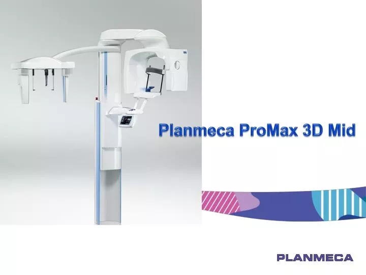
Planmeca ProMax 3D Mid
650 likes | 1.71k Views
Planmeca ProMax 3D Mid. Planmeca ProMax 3D models. Planmeca ProMax 3D family offers a solution for the most demanding imaging needs, producing various imaging sizes with one concept – an ideal imaging size for different maxillofacial applications. Cone beam 3D technology. Flat panel

Planmeca ProMax 3D Mid
E N D
Presentation Transcript
Planmeca ProMax 3D models • Planmeca ProMax 3D family offers a solution for the most demanding imaging needs, producing various imaging sizes with one concept – an ideal imaging size for different maxillofacial applications.
Cone beam 3D technology Flat panel detector Line detector object object Movement of translation and axis of rotation X-ray source X-ray source • Cone beam 3D X-ray device takes the whole 3 dimensional volume with wide conical beam in one scan instead of many slices with many scans.
3D technology • CBCT - Cone Beam Computed Tomography • CBVT - Cone Beam Volumetric Tomography • DVT – Digital Volume Tomography
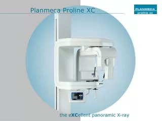
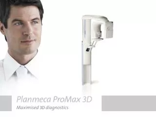
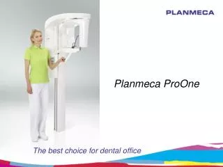
Planmeca ProMax 3D Mid • Real all in one X-ray • 3D imaging • ProFace 3D photo • SmartPan panoramic imaging system (optionally Dimax sensor) • Cephalometry • Simple and easy patient positioning • Robotic SCARA movement, adjustable volume place • Selectable image volume size • Latest flat panel technology • Pulsed X-ray generator
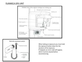
Easy positioning • Flexible head band patient support • Patient standing or sitting • Vertical fine-adjustment of the volume • Automatic vertically driving patient positioning system makes the stitching of basic volumes easy
3D image volume location • Pre-designed imaging programs for different targets • Dental programs: Incisors, Canines, Premolars, Molars, TMJ • ENT (Ear, Nose, Throat) programs: Sinus, Middle ear, Temporal bone, Vertebrae, Airways • SCARA-arm can position the rotation centre at any place • Adjustment of the volume diameter is possible • Fine adjustment with thumb wheel, joystick and laser lights • Exposure values automatically selected according to patient size and selected program
3D image volume sizes • From small Ø40 x 50 mm to large Ø160 x 160 mm • Ø70mm and smaller volumes can be freely placed in the imaging area and also horizontally stitched. • Maximum FOV (Ø160 x 160) can be reached with automatic vertical stitching program. • Programs match diagnostic task • Minimize radiated volume
Imaging modes • High resolution mode • Voxel size 100 µm • 400 or 450 frames • Effective exposure time 12 s • HD (High Definition) mode • Voxel size 150 µm • 500 or 600 frames • Effective exposure time 15 s • Normal resolution mode • Voxel size 200 µm • 400 or 450 frames • Effective exposure time 12 s • Low dose mode • Voxel size 400 or 600 µm • 300 or 450 frames • Lower exposure values • Effective exposure time 2.4 s • Remarkably lower dose • Small file size
3D Dental programs • Tooth • Adult: Ø40 x 50 mm, Ø40 x 70 mm, • Child: Ø34 x 42 mm Ø34 x 60 mm • Voxel Size: 100µm, 150 µm, 200µm, 400µm • Teeth • Adult: Ø70 x 50 mm, Ø70 x 70 mm, Ø90 x 50 mm, Ø90 x 90 mm • Child: Ø60 x 42 mm, Ø60 x 60 mm, Ø75 x 42 mm, Ø75 x 75 mm • Voxel Size: 150 µm, 200µm, 400µm • Jaw • Adult & child: Ø160 x 50 mm, Ø160 x 90 mm • Voxel Size: 200µm, 400µm, 600 µm
3D Dental programs • Face (stitched) • Adult & child: Ø160 mm x 160 mm • Voxel Size: 400µm, 600µm • Horizontal pair (stitched) • Adult: Ø40 x 50 mm, Ø40 x 70 mm, Ø70 x 50 mm, Ø70 x 70 mm • Child: Ø34 x 42 mm, Ø34 x 60 mm, Ø60 x 42 mm, Ø60 x 60 mm • Voxel Size: 400µm, 600µm
3D ENT programs • Sinus • Adult & child: Ø90 x 90 mm, Ø90 x160mm, Ø160 x 90 mm, Ø160 x 160 mm • Voxel Size: 200µm, 400µm, 600 µm • Middle ear • Adult: Ø40 x 50 mm, Ø70 x 70 mm • Child: Ø34 x 42 mm, Ø60 x 60 mm • Voxel Size: 100 µm, 150 µm, 200µm • Middle ear pair • Adult: Ø40 x 50 mm, Ø70 x 70 mm • Child: Ø34 x 42 mm, Ø60 x 60 mm • Voxel Size: 200 µm, 400 µm
3D ENT programs • Temporal bone • Adult: Ø70 x 70 mm • Child: Ø60 x 60 mm • Voxel Size: 150 µm, 200µm • Temporal bone pair • Adult: Ø70 x 70 mm • Child: Ø60 x 60 mm • Voxel Size: 200µm, 400 µm
3D ENT programs • Vertebrae • Adult: Ø70 x 70 mm • Child: Ø60 x 60 mm • Voxel Size: 200µm, 400µm • Airways • Adult: Ø70 x 70 mm • Child: Ø60 x 60 mm • Voxel Size: 200µm, 400 µm
3D programs • Impression scan • A special Impression Scan program produces very precise 3D images of impressions and plaster casts.
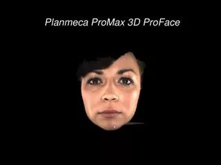
Planmeca ProMax 3D ProFace • The first CBVT unit integrated 3D surface scan • CBVT volume and 3D photo in one scan • 3D photo acquisition can be acquired separately • Totally radiation-free process
Planmeca ProMax 3D ProFace • Lasers scan face geometry • Digital cameras capture colourtexture • Software calculates information into a 3D photo • No additional equipment required; all components are integrated to sensor housing
SmartPan system • Images are taken with the same flat panel 3D sensor • Work flow is the same as in 2D imaging programs • Same patient positioning • Same image processing parameters • Frames grabbed with slit beam and narrow sensor area • Tomosynthesis applied to calculate the panoramic images
SmartPan system • ~2000 frames, snap shots, are taken during the panoramic scan • 9 different panoramic layers with 2 mm shift are calculated from the frames • +1 panoramic image in which the layer is optimised automatically
SmartPan system • 10 panoramic images are saved as a stack • You can browse the images and select the best
3D Mid optimized SmartPan Optimized SmartPan takes the panoramic image only from teeth area • Reduces the patient dose
3D Mid optimized SmartPan + TMJ TMJ’s can be imagined separately • Optimal imaging angle for TMJ’s • Images are save in a study in Romexis
Planmeca Romexis • 2D Imaging • 2D Implant Planning • 3D Imaging • 3D Explorer • 3D Cross Sections • 3D Implant Planning • 3D TMJ • 3D ProFace • Exporting and distributing images • Printing & Reporting • Patient & User Management • Radiology Module • Integration / Compatibility
Romexis 3D – Digital Cast & STL • Digital Cast tool & STL format export • Instant tool for converting ProMax 3D Impression Scans into digital dental models. • From 3D Impression Scan to STL dental model in less than 2 minutes! • Export 3D rendering surface in STL model directly from Romexis software • STL export especially designed for Digital Cast models • Use STL models in 3rd party software Negative Digital Cast STL model 3rd party software
Planmeca ProMax 3D Mid and cephalometry • Digital cephalostat is ideal for cephalometry • all information for orthodontic planning • lower radiation dose • faster procedure • 3D to be used for detailed e.g. occlusion views, impacted teeth etc. if needed • Same Promax unit for both studies • Optimum for orthodontic imaging
Cone beam 3D technology Flat panel detector axis of rotation object X-ray source Line detector Movement of translation and axis of rotation object X-ray source • Planmeca ProMax 3D works with cone beam computed tomography (CBCT) principle. • CBCT uses wide conical or pyramid shaped x-ray beam instead of narrow fan beam. • The 3D image is one 3-dimensional volume, not a pile of narrow slices. • CBCT scan is only one rotation around the patient. Medical CT needs several rounds around the patient. • CBCT: • is faster • has lower dose • has better resolution
3D Technology • Snap shot images taken with synchronized X-ray pulses • Stroboscopic effect for maximum clarity of the images • Accumulated X-ray exposure time is 2.8 – 18s – reduced dose • Proprietary 3D reconstruction algorithm • Voxel size 100, 200 or 400 µm depending on resolution mode and volume size • Resolution up to 5 lp/mm (Nyquist)
ProMax 3D Mid Scanning • Symmetric scanning • C-ram rotates • Magnification 1.8x • Scan angle 210° • 300 - 500 frames • Max. volume Ø70 x 70 mm • Asymmetric off-set scanning • Elbow arm rotates • Magnification 1.44x • Scan angle 360 deg • 450 - 600 frames • Max. volume Ø160 x 90 mm
Asymmetric off-set scanning • Sensor shift changes the acquisition geometry and reduces the final image quality • The shift of whole c-arm keeps the acquisition geometry constant and produces better final image SCARA!
Isotropic voxel • CBCT has always an isotropic voxel • The reconstruction can produce any size of voxel • The voxel is always perfect cube • The measurements are exact • Voxel size is typically 0.1 – 0.5 mm • CT has an anisotropic voxel • The voxel is always a “brick” • The pitch (= distance between spiral rounds = layer thickness) varies and causes distortion in the 3D measurements. • The layer thickness is typically 0.5 – 0.8 mm
3D Technology • Pulsed X-ray produces sharp images with less dose.
3D Technology – Flat Panel • Planmeca ProMax 3D flat panel imaging chain • Conventional imaging chainwith Image Intensifier X-ray Tube – Patient – Flat Panel - Digital Image X-ray Tube – Patient – Image Intensifier – TV Camera – Digital Image Modern Flat Panel Technology for maximum performance
3D Technology – Flat Panel • Image intensifier has both distortion and brightness non-uniformity which is absent from the flat panel detector • Image intensifier needs periodical maintenance. It has limited life span 3-6 years. • It is sensitive to magnetic or electrical fields. • It is over 60 years old technology.
Flat Panel vs Image Intensifier • Image intensifier image quality decreases sharply over time. Flat panel’s life time is longer.
3D technology – Tube Current Modulation less more • Different attenuation properties across and along the patient's head • Tube current (mAs) can be dynamically adjusted • Reduces patient dose and improves image quality
Radiation dose • Modern low dose medical CT: 685 - 1400 µSv (ICRP 2008) • Radiation dose of CBCT: 20 - 1000 µSv • ProMax 3D: 28 - 122 µSv • Panoramic image 3 – 23 µSv • ProMax pan: 10 – 23 µSv • ProMax SmartPan: 12.5 – 30 µSv • Intraoral image <8 µSv
The End • More information: • Erkki Hiltunen • Product Manager, X-rays • tel: +358 20 7795 456 • erkki.hiltunen@planmeca.com • Mark Niemi • Product Manager, X-rays • tel: +358 20 7795 743 • mark.niemi@planmeca.com • More information: • Osku Sundqvist • Product Manager, Software • tel: +358 20 7795 793 • osku.sundqvist@planmeca.com 3/2012