Download
1 / 1
10 likes | 129 Views
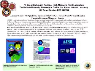
PI: Greg Boebinger, National High Magnetic Field Laboratory Florida State University, University of Florida, Los Alamos National Laboratory NSF Award Number: DMR-0084173. A Comprehensive 3D Digital Atlas Database of the C57BL/6J Mouse Brain Developed Based on

E N D
PI: Greg Boebinger, National High Magnetic Field LaboratoryFlorida State University, University of Florida, Los Alamos National Laboratory NSF Award Number: DMR-0084173 A Comprehensive 3D Digital Atlas Database of the C57BL/6J Mouse Brain Developed Based on Magnetic Resonance Microscopy Images: AMRIS investigators published more than 30 peer reviewed papers in 2005, including 2 additional cover articles and a feature in Science. The use of transgenic mice has revolutionized biomedical research and genetics, and numerous genetic diseases can be studied and better understood with a “mouse model” of the disease. Before the effects of the mutations can be fully understood, however, a bench-mark study of a normal mouse is needed. Drs. Blackband and Grant at UF and Dr. Benveniste at Brookhaven National Laboratory have used microimaging at 750 MHz in AMRIS to make a comprehensive 3D digital mouse brain atlas of the standard lab mouse, C57BL/6J [Ma, et al., Neuroscience, 135, 1203-15 (2005)]. ThisDr. Mareci’s laboratory at UF has used very high-resolution imaging of spinal cord injury and compared in vitro MRI, in vivo MRI, and standard histology. Berens, et al., Am. J. Neuroradiol. 1612-22 (2005)]. This work was also recognized by a cover article in the prestigious American Journal of Neuroradiology.