Download
1 / 17
180 likes | 243 Views
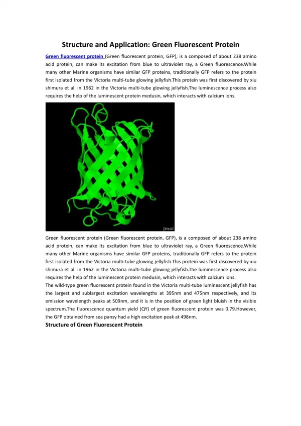
Whole-body optical imaging of green fluorescent protein-expressing tumors and metastases.

E N D
Whole-body optical imaging of green fluorescent protein-expressing tumors and metastases Meng Yang*, , , Eugene Baranov*, Ping Jiang*, Fang-Xian Sun*, Xiao-Ming Li*, Lingna Li*, Satoshi Hasegawa*, , , Michael Bouvet , Maraya Al-Tuwaijri*, , Takashi Chishima*, , , Hiroshi Shimada , A. R. Moossa , Sheldon Penman§, and Robert M. Hoffman*, ,¶
Stable, high-level GFP-expressing murine melanoma transductants in vitro
External images of murine melanoma (B16F0-GFP) metastasis in brain. Murine melanoma metastases in the mouse brain were imaged by GFP expression under fluorescence microscopy. Clear images of metastatic lesions in the brain can be visualized through the scalp and skull
External images of B16F0-GFP bone metastasis. In the proximal tibia of the left hind leg of C57BL/6 mouse (hair removed). No metastasis can be detected under bright-field microscopy (A). Clear, external images of metastatic lesions of B16F0-GFP in the proximal tibia of the intact mouse were obtained under fluorescence microscopy (B). Time course metastatic growth of B16F0-GFP in the proximal tibia of the intact nude mouse was imaged externally under fluorescence microscopy (C-E).
External images of B16F0-GFP colonizing the liver. A metastatic lesion of B16F0-GFP in the liver growing at a depth of 0.8 mm after portal vein injection was externally imaged through the abdominal wall of the intact nude mouse
External and internal images of liver lesions of AC3488-GFP. (A) Lateral, whole-body image of metastatic liver lesions of a GFP-expressing human colon cancer in the left (thick arrow) and right lobes (fine arrow) of a live nude mouse at day 21 after surgical orthotopic transplantation. (B) Cross-section of mouse shown in A corresponding to the level of the external image of the tumor in the liver that was acquired (A).
External and internal images of bone metastasis of AC3488 GFP. External fluorescent whole-body images compared with direct images of skeletal metastases. (A) External images of tumors in the skeletal system including the skull (arrow heads), scapula (thick arrows), spine (fine arrows), and liver metastasis (largest arrows) in a dorsal view of live,
Table 1. Minimum-sized fluorescent optical tumour images at increasing depth
Visualizing gene expression by whole-body fluorescence imaging Meng Yang*, Eugene Baranov*, A. R. Moossa , Sheldon Penman , and Robert M. Hoffman*, ,§ * AntiCancer, Inc., 7917 Ostrow Street, San Diego, CA 92111; Department of Surgery, University of California, 200 West Arbor Drive, San Diego, CA 92103-8220; and Department of Biology, Massachusetts Institute of Technology, 77 Massachusetts Avenue, Cambridge, MA 02139-4307 • Contributed by Sheldon Penman, August 14, 2000
Materials and Methods • Animals. Four- to 6-week-old both male and female nude/nude, nude/+, and C57BL/6 mice were used. All animal studies were conducted in accordance with the principles and procedures outlined in the National Institutes of Health Guide for the Care and Use of Animals under assurance number A3873-1. Mice were fed with autoclaved laboratory rodent diet (Tecklad LM-485, Western Research Products, Orange, CA). • DNA Expression Vector. The adenoviral (vAd) vector AdCMV5GFPAE1/AE3 [vAd-green fluorescent protein (GFP)] (Quantum, Montreal, Canada) expresses enhanced GFP and the ampicillin resistance gene. • Delivery of vAd-GFP to Various Organs.Brain. The parietal bone of the skull was exposed after an upper midline scalp incision. Twenty microliters containing 8 × 10¹⁰ plaque-forming units (pfu)/ml vAd-GFP per mouse was injected in the brain by using a 1-ml 27G1/2 latex-free syringe (Becton Dickinson). The puncture hole in the skull was plugged with bone wax. The incision in the scalp was closed with a 7-0 surgical suture in one layer. The animals were kept under isofluorane anesthesia during surgery
Fig. 1. External and internal images of vAd-GFP gene expression in various organs. External fluorescent whole-body images were compared with direct images of vAd-GFP gene expression in various organs. Series of external fluorescence images of vAd-GFP gene expression in brain, liver, pancreas, prostate, and tibia, (A, C, E, G, and I) compared with corresponding images of the exposed organs (B, D, F, H, and J).
Fig. 2. External whole-body image of vAd-GFP gene expression in the brain. An external image of vAd-GFP gene expression in the brain acquired from a nude mouse in the light box 24 h after gene delivery
from a series of whole-body images determined that vAd-GFP expression could be first visualized by 5 h, 15 min after local introduction of vAd-GFP gene in a nude mouse. Increases in expression could be visualized in 30-min intervals.
Expression [I'GFP] could be first visualized in the liver by 7 h, 45 min after introduction of vAd-GFP gene in a nude mouse. Increases in expression could be visualized in 30-min intervals
An external External whole-body image of vAd-GFP gene expression in the liver. image of vAd-GFP gene expression acquired from a nude mouse in the light box 72 h after gene delivery. Lateral, whole-body image of transgene expression in the liver can be clearly visualized through the abdominal wall
T50 only T50Luc right ear T50 Luc right and left T50Luc no ear C3H/T50 LUC 0.2x10e6 cells Day 22 after inoculation.
Bladder Luc adsv40 No Luc bladder adsv40 overlay bio open rat Open rat overlay Rat no bladder Bio bladder overlay Bladder only no bladder Overlay adsv40Luc. WISTAR bladder (catheter) 45 Hrs