Download
1 / 17
3.15k likes | 10.86k Views
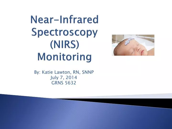
Near-Infrared Spectroscopy (NIRS) Monitoring. By: Katie Lawton, RN, SNNP July 7, 2014 GRNS 5632. Objectives. To understand what NIRS is To understand how NIRS works To become acquainted with the NIRS machine and where to place the probe

E N D
Near-Infrared Spectroscopy(NIRS)Monitoring By: Katie Lawton, RN, SNNP July 7, 2014 GRNS 5632
Objectives • To understand what NIRS is • To understand how NIRS works • To become acquainted with the NIRS machine and where to place the probe • To understand how NIRS can be utilized in the clinical setting • To understand the beneficial significance of NIRS
Objectives • To understand the relationship between cerebral and somatic regional oxygen saturations • To become familiar with normal cerebral and somatic rSO2 ranges and interventions to improve rSO2 • To provide research regarding the use of NIRS in the clinical setting in relation with patient outcomes
What is NIRS? • Noninvasive, continuous tissue oxygenation monitoring • Measures concentrations of oxyhemoglobin and deoxyhemoglobin • Used to measure regional oxygen saturation (rSO2) • Cerebral and Somatic • Can be to measure oxygenation in: • Brain • Renal • Liver • Extremities • Commonly used during cardiac surgery and emergency situations (Covidien, 2011)
Factors That Affect Cerebral Oxygen Saturation • Increase • Rise in oxygen delivery • Diminished oxygen demand • Decrease • Decrease in oxygen delivery • Uncompensated rise in demand (Covidien, n.d.)
Why is NIRS used at the bedside? • Helps in early identification of complications associated with • Low cardiac output • Shock and seizures • Renal failure • Neurological damage
Markers of Perfusion (Covidien, n.d.)
How does it work? • Measure the relationship between light and concentration of the compound • Evaluates the transparency of tissues to the near infrared light to determine tissue oxygenation • Uses Beer-Lambert equation: log (I/Io) = L C I: measures power of light at the detector after it passes through the tissue Io: measured power of light at the emitter before it enters the tissue L: path length of the light from the emitter to detector C: concentration of the absorbing compound in the tissue (Kurth, 2006)
NIRS Equipment Spectrophotometer I/Io Probe
How is the rSO2 determined by the probe placement? • The probe (I/Io) is placed on the on the skull (cuvette) • LED light is emitted in tissue (scalp, skull and brain) • Distal and proximal detector (on other end of probe) provide information regarding the oxygenation of tissues after light is emitted through the cuvette and relayed to the detector (Covidien, 2011)
NIRS Monitor • L: Left Cerebral • R: Right Cerebral • S: Somatic (Covidien, n.d.)
Probe Placement • Cerebral Probe Placement • L/R side of forehead • Somatic • Renal area • Abdomen • Upper extremites • (arm) • Lower extremities • (calf, thigh) (Covidien, n.d.)
RelatonshipBetween Cerebral & Somatic Regions • Cerebral • High flow/ high extraction organ • Compensatory mechanisms • Autoregulation • Flow metabolism coupling • If autoregulation is intact, cerebral desaturations are a late warning of shock • Somatic • Variable flow/ low O2 extraction • Flow influenced by sympathetic tone • Somatic desaturations are early sign of shock due to compensatory mechanisms (Covidien, n.d.)
Cerebral Targets • Provides indication of hypoxia and cerebral perfusion • rSO2 range 60-80% • During cardiac surgery a drop in rSO2 <45% or a 25% drop from the individual baseline is critical • Studies have shown a correlation between rSO2 in cardiac surgery and postoperative outcome • Intraoperativedesatuartion associated with cognitive dysfunction, stroke and increased length of stay with decreased rSO2 (Scheeren, Schober & Schwarte, 2012)
Somatic Targets • Lower oxygen usage than cerebral • rSO2 range: 5-20 higher than cerebral rSO2 • Changes in variance may indicate pathology (Covidien, n.d.)
Interventions to Improve rSO2 • Cerebral • Increase cerebral perfusion pressure • Increase arterial oxygen content • Decrease cerebral vascular resistance • Decrease cerebral metabolic rate • Somatic • Increase cardiac output • Reduce sympathetic outflow • Increase Hct • Avoid hypothermia/hyperthermia • Regional vasodilation (Covidien, n.d.)
References Covidien. (n.d). INVOS Cerebral/Somatic Oximeter: Quick reference guide for pediatric use. Retrieved from https://lane.stanford.edu/portals/cvicu/HCP_Equipment/NIRS-INVOS_Reference_Guide.pdf Covidien. (2011). INVOS Cerebral/Somatic Oximeter. Retrieved from http://www.covidien.com/rms/imageServer.aspx?contentID=27565&contenttype=application/p df&originalFileName=1.3.3.1_Neonatal%20Brochure.pdf Hill, L. (n.d.). Cerebral blood flow and intracranial pressure. Retrieved from http://www.frca.co.uk/Documents/17 0907%20Cerebral%20physiology%20I.pdf Kurth. (2006). Near infrared spectroscopy. Retrieved from http://www.pedsanesthesia.org/meetings/2006winter/pdfs/Friday_Kurth.pdf Marimon, G.A., Dockery, W.K. Sheridan, M. J. & Agarwal, S. (2012). Near-infrared spectroscopy cerebral and somatic (renal) oxygen saturation correlation to continuous venous oxygen saturation via intravenous oximetry catheter. Journal of Critical Care, 27, 314.e13-314.e18. doi: 10.1016/j.jcrc.2011.10.002 Scheeren, T.W.L, Schober, P. & Schwarte, L.A. (2012). Monitoring tissue oxygenation by near infrared spectroscopy: background and current applications. Journal of Clinical Monitoring and Computing, 26 (4), 279-287. doi: 10.1007/s10877-012-9348-y