
Development of Myelofibrosis-Like Disease in JAK2V617F Bone Marrow Transplantation Mice
This study explores the development of myelofibrosis-like disease in mice undergoing bone marrow transplantation (BMT) using JAK2V617F-expressing donor cells. BALB/c mice were transplanted with either JAK2V617F or JAK2WT cells, and assessments were made 80 days post-BMT. Key hematological parameters such as white blood cells, red blood cells, reticulocytes, and platelets were evaluated. The effects of NS-018 on erythroid colony formation were analyzed in JAK2V617F transgenic mice, and phosphorylation status of Src in treated cells was assessed. Results provide insights into the pathophysiology of myelofibrosis.

Development of Myelofibrosis-Like Disease in JAK2V617F Bone Marrow Transplantation Mice
E N D
Presentation Transcript
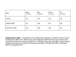
Supplementary Table 1. Development of myelofibrosis-like disease in JAK2V617F bone marrow transplantation (BMT) mice. BALB/c mice were subjected to BMT with bone marrow donor cells retrovirally transduced to express JAK2V617F or JAK2WT. Eighty days after BMT, peripheral blood samples and organs were collected. White blood cell (WBC), red blood cell (RBC), reticulocyte (RETIC) and platelet (PLT).
a b c d Supplementary Figure 1. X-ray structures of (a) NS-018, (b) AZD1480, (c) CP-690550, and (d) AT9283 in complex with JAK2 kinase. Protein Data bank IDs for AZD1480, CP-690550, and AT9283 are 2XA4, 3FUP, and 2W1I, respectively.
Supplementary Figure 2. Effect of NS-018 on erythroid colony formation in JAK2V617F transgenic mice. The generation and genotyping of JAK2V617F transgenic mice were carried out as described previously 15. Bone-marrow cells were harvested by flushing the femurs and tibias of JAK2V617F transgenic mice or WT control mice with phosphate-buffered saline. A total of 2 × 10⁵ cells were treated with increasing concentrations of NS-018 in MethoCult M3334 methylcellulose medium (StemCell Technologies, Vancouver, BC, Canada) in the presence of 3 U/ml erythropoietin. Experiments were performed in triplicate. Erythroid colony-forming units (CFU-E) were counted on day 3 and a two-way factorial analysis of variance was performed with SAS version 9.1.3 (*P<0.001). Bars represent the mean ± s.e.m. (n = 4).
Supplementary Figure 3. Phosphorylation status of Src in Ba/F3-JAK2V617F cells. Ba/F3-JAK2V617F cells were treated with increasing concentrations of NS-018 or ruxolitinib for 3 h. Total cell lysate was subjected to SDS-PAGE, transferred to a PVDF membrane and probed with antibody specific for phospho-Src family (Tyr416) or Src (Cell Signaling Technology).