Download

1 / 57
570 likes | 1.06k Views
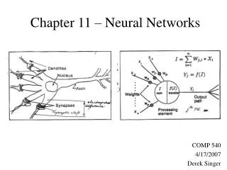
Chapter 39 Neural Signaling and Chapter 40 Neural Regulation. The Nervous System. Parts of a Neuron. Receive stimuli Produce and transmit electrical signals ( aka nerve impulses/action potentials) Synthesize and release neurotransmitters Draw neuron Many axons make nerve

E N D
Chapter 39 Neural Signaling and Chapter 40 Neural Regulation The Nervous System
Parts of a Neuron • Receive stimuli • Produce and transmit electrical signals ( aka nerve impulses/action potentials) • Synthesize and release neurotransmitters • Draw neuron • Many axons make nerve • Tracts/pathways – bundles of axons in CNS • Ganglia – groups of cell bodies outside CNS • Nuclei – groups of cell bodies inside CNS
Fig. 48-12 Node of Ranvier Layers of myelin Axon Schwann cell Schwann cell Nucleus of Schwann cell Nodes of Ranvier Axon Myelin sheath 0.1 µm
Neural Signaling: 4 processes(communication among neurons) • Reception • Detect a stimulus • Neurons and sense organs • Transmission • Message sent along neuron, between neurons, to effector • Integration • Sort and interpret incoming info, determine response • Action by effectors • Actual response to stimulus
Fig. 48-3 Sensory input Integration Sensor Motor output Central nervous system (CNS) Effector Peripheral nervous system (PNS)
Types of Neurons • Sensory neurons aka Afferent neurons • Info TO CNS • Interneurons aka Association neurons • Info from afferent neurons to interneurons • Integrate and output • Most common • Cell body and axon in CNS • Motor neurons aka Efferent neurons • Carries message from CNS to effector
Glial Cells (neuroglia)– support and protect neurons, regulatory functions - CNS • Microglia • Phagocytes – remove debris • Astrocytes • Star-shaped • Provide glucose to neurons • Regulate extracellular fluid • Oligodendrocytes • Form sheath of myelin around neurons • Schwann cells • Outside of CNS • Form sheaths around some axons
Fig. 49-6 PNS CNS Neuron VENTRICLE Astrocyte Ependy- mal cell Oligodendrocyte Schwann cells Microglial cell Capillary (a) Glia in vertebrates 50 µm (b) Astrocytes (LM)
Nerve impulse • Myelin • Multiple sclerosis
Synapses • Presynaptic neuron / Postsynaptic neuron • Electrical synapse • Chemical synapse
Electrical synapse • 2 neurons very close together • Interiors of 2 cells physically connected by protein channel • Ion passage between cells, permitting an impulse to be directly and rapidly transmitted from pre to postsynaptic neuron • Used for escape responses
Chemical synapse • More common • 2 neurons separated by synaptic cleft • Depolarization of property of PM so when action potential reaches end of axon it is unable to jump the gap • Electrical signal must be converted to chemical signal (neurotransmitter) • When postsynaptic neuron reaches threshold depolarization, it transmits an action potential
Fig. 48-15 5 Na+ K+ Synaptic vesicles containing neurotransmitter Presynaptic membrane Voltage-gated Ca2+ channel Postsynaptic membrane Ca2+ 1 4 6 2 3 Synaptic cleft Ligand-gated ion channels
Neurotransmitter – conduct neural signal across synapse and bind to chemically activated ion channels in PM of postsynaptic neuron • Ex: acetycholine • Norepinephrine, serotonin, dopamine • Neuromodulator – messengers that modify the effects of specific neurotransmitters • Some amplify/dampen response by postsynaptic cell
How Neurotransmitters (NT) work • Stored in synaptic terminals in synaptic vesicles • Action potential reaches synaptic terminal • Voltage-gated calcium channels open • Calcium ions from extracellular fluid flow into synaptic terminal • Ca ions cause synaptic vesicles to fuse with presynaptic membrane and release NT into synaptic cleft by exocytosis • NT diffuse across synaptic cleft and combine with specific receptors on dendrites or cell bodies of postsynaptic neurons (or PM of effector cells)
Ligand-gated ion channel – NT receptor, chemically activated • Ligand (NT) binds with receptor and ion channel opens • Ex: Ach receptor is ion channel for passage of Na+ and K+
Repolarization - Quick • Excess NT must be removed • Degraded by enzymes • Ex: Acetylcholinesterase breaks Ach choline + acetate • Active transport back into synaptic terminal = reuptake • Repackaged and recycled
Drugs inhibit reuptake • Antidepressants • SSRIs – selective serotonin reuptake inhibitors • Fluoxetine (Prozac) • Cocaine - dopamine
NT – different effects with different neurons • Ach • Excite – skeletal muscle • Inhibit – cardiac muscle • Excitatory postsynaptic potential (EPSP) • Change in membrane potential that brings neuron closer to firing • Inhibitory postsynaptic potential (IPSP) • Change in membrane potential that takes the neuron farther away from firing
Chapter 40: Neural Regulation • Vertebrate Nervous System • CNS • Complex brain continuous with spinal cord • Central control • Integrate incoming info • Determine appropriate response • PNS • Sensory receptors and nerves (communication lines)
Fig. 49-4 Peripheral nervous system (PNS) Central nervous system (CNS) Brain Cranial nerves Spinal cord Ganglia outside CNS Spinal nerves
Fig. 49-5 Gray matter White matter Ventricles
Cranial nerves • Link body parts to brain • Spinal nerves • Link body parts to spinal cord
Vertebrate Brain • Brainstem = medulla, pons, midbrain • Medulla • Most posterior • Regulate respiration, heartbeat, BP, swallowing, coughing, vomiting • Pons • Mammals • Bulge anterior of brain stem • Bridge – connects spinal cord and medulla with upper parts of brain • Regular respiration • Relay impulses from cerebrum cerebellum
midbrain • (mesencephalon) • Visual reflexes (pupil constriction) • Auditory reflexes • Muscle tone and posture
Cerebellum • Muscle activity – tone, posture, equilibrium (balance) • Thalamus • Relay center for motor and sensory messages
Hypothalamus • Below thalamus • Olfactory centers • Principal integration center for regulation of viscera • Provides input to medulla and spinal cord that regulate heart rate, respiration, digestive function • Controls body temp. • Regulates appetite, water balance • Emotional/sexual responses • Links nervous and endocrine systems, produces certain hormones
Cerebrum • Most prominent • Olfactory • R and L hemispheres • Mostly white matter (mainly myelinated axons that connect various parts of brain) • Surface convolutions = numerous folds • Expands surface • Sulci – furrows between convolutions if shallow • Fissures if deep • # folds associated with complexity of brain function
Gray matter – cerebral cortex • Makes up outer portion of cerebrum • Contains cell bodies and dendrites
Human CNS • Well-protected brain and spinal cord • 3 layers connective tissue (meninges) and encased in bone • 3 meningeal layers • Outer dura mater • Middle arachnoid • Thin vascular pia mater (adheres closely to tissue of brain and spinal cord)
Meningitis – disease where these coverings become infected and inflamed • Cerebrospinal fluid (CSF) • Between arachnoid and pia mater, in subarachnoid space • Produced by choroid plexus = special networks of capillaries extend up from pia mater into brain ventricles; extract nutrients from blood and adds them to CSF • Choroid plexus and arachnoid serve as barrier between blood and CSF (prevent harmful substances from entering the brain)
CSF • Shock absorber • Cushions brain and spinal cord • Medium for exchange of nutrients and waste products between brain and blood
Spinal Cord • Base of brain to L2 vertebra • Central canal surrounded by gray matter (cell bodies, dendrites, unmyelinated axons, glial cells) in “H” shape • White matter (outside gray matter) of myelinated axons in bundles (tracts)
Reflex action • Relatively fixed response pattern to a simple stimulus • Predictable, automatic, unconscious • Ex: breathing
Withdrawal reflex • Touch hot stove, jerk hand away • Route of the message • Pain receptor in skin sensory neuron spinal cord association neuron appropriate motor neuron group of muscles • Same time – message sent to conscious areas of brain (up spinal cord) • Aware, feel pain (not part of reflex)
Fig. 49-3 Cell body of sensory neuron in dorsal root ganglion Gray matter Quadriceps muscle White matter Hamstring muscle Spinal cord (cross section) Sensory neuron Motor neuron Interneuron
Human Cerebrum • Cerebral cortex (outer part of cerebrum) • R and L cerebral hemispheres • Functionally divided into 3 area • 1 – sensory – receive incoming signal from sense organs • 2 – motor – control voluntary movement • 3 – association – link sensory and motor areas, responsible for thought, learning, language, memory, judgment, personality
Occipital lobes – vision • Temporal lobes – hearing • Central sulcus – groove across top of each hemisphere from medial to lateral edge • Partially separate frontal lobes from parietal lobes • frontal lobes – skeletal muscles • Parietal lobes – heat, cold, touch, pressure from skin
Fig. 49-15 Frontal lobe Parietal lobe Somatosensory cortex Motor cortex Somatosensory association area Speech Frontal association area Taste Reading Speech Hearing Visual association area Smell Auditory association area Vision Temporal lobe Occipital lobe
Size of motor area in brain for a given body part proportional to complexity of movement involved • Ex: hands and face = large areas • One side of brain controls opposite side of body • Uppermost part of cortex controls lower limbs of body
White matter of cerebrum under cerebral cortex • Nerve fibers of white matter connect the cortical areas with 1 another and with other parts of the nervous system
Corpus callosum - Large band of white matter, connects R and L hemispheres • Basal ganglia • Deep in white matter • Paired groups of nuclei (gray matter) • Coordination and movement • Send signals to midbrain
Cerebral cortex – integrates info about diverse activities • Arousal, sleep, emotion, information processing
The Brain and Sleep-wake • “brain waves” / electrical potentials generated by active neurons can be measured • Recorded by electroencephalogram (EEG) • Electrodes taped to scalp and activity of cerebral cortex measured
Alpha waves • Most regular indication of activity • Occur rhythmically ~ 10/sec. • Mostly from visual areas in occipital lobe (rest quietly, eyes closed) • Beta waves • Rapid, irregular waves, eyes open • Fast frequency • Heightened mental activity • Delta and theta waves • Slow, large waves associated with certain stages of sleep
Fig. 49-11 Key Low-frequency waves characteristic of sleep High-frequency waves characteristic of wakefulness Time: 1 hour Location Time: 0 hours Left hemisphere Right hemisphere
Learning • Process by which we acquire info as a result of experience • Memory • Process by which information is encoded, stored, retrieved