Download
1 / 33
340 likes | 547 Views
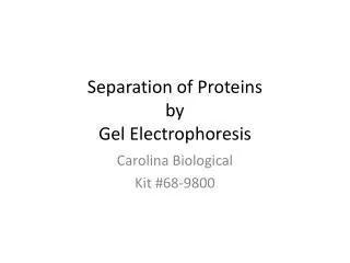
Separation of Proteins See Reference Textbook of biochemistry with clinical correlations. Sixth edition. Thomas M. Devlin. Page117-120. MALIK ALQUB MD. PHD. Separation of different cell compartements. Differential centrifugation

E N D
Separation of ProteinsSee Reference Textbook of biochemistry with clinical correlations.Sixth edition. Thomas M. Devlin. Page117-120 MALIK ALQUB MD. PHD.
Separation of differentcellcompartements • Differential centrifugation • Differential centrifugation is used to separate certain organelles from whole cells for further analysis of specific parts of cells. In the process, a tissue sample is first homogenised to break the cell membranes and mix up the cell contents. The homogenate is then subjected to repeated centrifugations, each time removing the pellet and increasing the centrifugal force.
Differential centrifugation • Differential centrifugation can separate cell compartments depending on their density against sucrose gradient • Whole cells and nuclei; • Mitochondria, lysosomes and peroxisomes; • Microsomes (vesicles of disrupted endoplasmic reticulum); and • Ribosomes and cytosol.
Separating Proteins from Mixtures • After separation of cell compartments • Proteins can be separated from each other based on differences in physical properties • Due to different amino acid sequences proteins differ in solubility, size, charge, and binding affinity and can be separated on either of these properties
Separation of proteinsupontheir • Charge • Size • Binding affinity
Separation of proteinsupontheir • Charge • Size • Binding affinity
Charge • Three main separtaionmethods • Chromatography • Isoelectricfocusing • Electrophoresis
Charge • Three main separtaionmethods • Chromatography • Isoelectricfocusing • Electrophoresis
An introduction to liquid chromatography • Protein solution applied to a column • Column = solid porous matrix (stationary phase) + liquid (mobile phase) • Proteins separated based on differing interactions with stationary and mobile phases • Mobile phase conditions can be adjusted to increase or decrease affinity of protein for stationary phase (gradient)
Fractionation during chromatography Proteins separated by chromatography are collected in fractions to keep them separated
His: 6.0 Glu: 4.1 Arg: 12.5 N-terminal amine: 8.0 C-terminal acid: 3.1 For this peptide: pI=pKa/N= 6.3 Positively charged at pH < 6.3 Negatively charged at pH > 6.3 Separation on the Basis of Charge H2N- Met Ala Asn Cys His Glu Ser Thr Glu Arg-COOH • All proteins are charged. Their charges depend on the relative number of acidic and basic amino acids in their primary structure. • All proteins have a pH value where they are uncharged: the isolelectic point (pI)
-- -- - - - -- - + + + -- -- + -- + + - - + + + - - - - + -- + -- -- -- + -- + - - - Ion Exchange Chromatography: • Positive or negatively charged resin can be used for separation of positive or negatively charged proteins • Sample of proteins is added • Washed with buffer to remove non specifically bound protein • Elute with increasing concentration of salt • Proteins with highest net charge come at last
Charge • Three main separtaionmethods • Chromatography • Isoelectricfocusing • Electrophoresis
Isoelectric Focusing • Isoelectric focusing methods are widely applied for the separation of proteins, peptides and enzymes. The principle: In a pH gradient the sample components migrate towards the anode or the cathode to the pH values, where their net charges are zero.
Charge • Threesepartaionmethods (not exclusive) • Chromatography • Isoelectricfocusing • Electrophoresis
Electrophoresis of Proteins Electrophoresis Electro - energy of electricity Phoresis - from the Greek, ‘phoros’ - to carry across • protein electrophoresis on cellulose acetate Principles • The principle of electrophoresis is based on the fact that a charged particle placed in an electrical field will migrate toward one of the electrodes of the field depending on the • electrical charge on the particle • size of the particle • strength of the electrical field • nature of the medium used to support the particle during the migration process
Separation of proteinsupontheir • Charge • Size • Binding affinity
Size • Threesepartaionmethods (not exclusive) • Gel Permeation Chromatography • SDS poly acrylamide gel electrophoresis
Size • Threesepartaionmethods (not exclusive) • Gel Permeation Chromatography • SDS poly acrylamide gel electrophoresis
UV time Gel Permeation Chromatography: Separating on Basis of Size Mixture of proteins • A mixture of proteins in a small volume is applied to a column filled with porous beads • Because large proteins cannot enter the beads, they emerge sooner than do small ones • A detector (e.g. UV) is used to detect protein fragments • Fragments are collected separately
Size • Threesepartaionmethods (not exclusive) • Gel Permeation Chromatography • SDS poly acrylamide gel electrophoresis
SDS poly acrylamide gel electrophoresis Protein is dissolved in detergent solution of SDS (sodium dodecylsulfate), a negatively charged surfactant that complexes with protein molecules Proteins denature and obtain a negative charge (1 SDS per 2 amino acids) Charge is now a proportional to the protein size Small proteins move faster through the gel matrix
Immunodetection Transfer of proteins from the gel to a nitrocellulose paper Immunodetection
Separation of proteinsupontheir • Charge • Size • Binding affinity
X X X X X X X X X X X X Affinity Chromatography: Separating on the Basis of Affinity • To separate proteins that recognize • a chemical group “X” • X is covalently attached to beads that are packed in a column • Sample of proteins is added • Washed with buffer to remove non specifically bound protein • X= receptors, antibody...etc
Summary of Protein Isolation & Purification • One of the main aims of biochemistry is to characterize and catalogue all proteins in the cell • We have discussed some important tools for separating proteins based on physical properties such as size, affinity, charge • Chromotography methods: ion exchange, affinity, gel permeation chromatography • Electrophoresis: isoelectric focusing, SDS PAGE,
Separation of ProteinsSee Reference Textbook of biochemistry with clinical correlations.Sixth edition. Thomas M. Devlin. Page117-120