Download
1 / 50
501 likes | 525 Views
This chapter delves into the complexities of human inheritance, exploring patterns of autosomal dominant and recessive traits, X-linked inheritance, and disorders such as Huntington Disease and Galactosemia. The role of sex chromosomes, karyotype preparation, and chromosomal abnormalities like aneuploidy and polyploidy are elucidated, offering a comprehensive view of genetic variation and its impact on human health. Dive into the intricacies of chromosome structure and inheritance for a deeper understanding of genetic diversity.

E N D
Separate Events • Each “event” is separate. The “history” does not necesarilly indicate what will happen. • Families with mostly girls or boys
Genes Fig. 12-2, p.188
Sex Chromosomes • Discovered in late 1800s • Mammals, fruit flies • XX is female, XY is male • XY is male, XX female • Human X and Y chromosomes function as homologues during meiosis
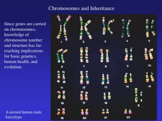
Karyotype Preparation Fig. 12-4, p.189
Karyotype Preparation • Arrested cells are broken open • Metaphase chromosomes are fixed and stained • Chromosomes are photographed through microscope • Photograph of chromosomes is cut up and arranged to form karyotype diagram
Karyotype Preparation Fig. 12-3a-e, p.189
Karyotype Diagram Fig. 12-3f, p.189
Autosomal Dominant Inheritance Trait typically appears in every generation Fig. 12-10a, p. 190
Autosomal Dominant Inheritance Fig. 12-5, p.190
Huntington Disorder • Autosomal dominant allele • Causes involuntary movements, nervous system deterioration, death • Symptoms don’t usually show up until person is past age 30 • People often pass allele on before they know they have it
Autosomal Recessive Inheritance Patterns • If parents are both heterozygous, child will have a 25% chance of being affected Fig. 12-10b, p. 191
enzyme 3 enzyme 1 enzyme 2 galactose-1- phosphate galactose-1- phosphate lactose galactose +glucose intermediate in glycolysis Galactosemia • Caused by autosomal recessive allele • Gene specifies a mutant enzyme in the pathway that breaks down lactose
Hutchinson-Gilford Progeria Fig. 12-7, p.191
The Y Chromosome • Fewer than two dozen genes identified • One is the master gene for male sex determination • SRY gene (sex-determining region of Y) • SRY present, testes form • SRY absent, ovaries form
The X Chromosome • Carries more than 2,300 genes • Most genes deal with nonsexual traits • Genes on X chromosome can be expressed in both males and females
x x Sex Determination diploid germ cells in female diploid germ cells in male meiosis, gamete formation in both female and male: eggs sperm X Y X X Fertilization: X X X XX XX sex chromosome combinations possible in new individual XY XY X Fig. 12-8a, p.192
At seven weeks, appearance of structures that will give rise to external genitalia At seven weeks, appearance of “uncommitted” duct system of embryo Effect of YChromosome Y chromosome present Y chromosome absent Y chromosome present Y chromosome absent testes ovaries 10 weeks 10 weeks ovary vaginal opening penis uterus vagina penis testis birth approaching Fig. 12-8bc, p.192
X-Linked Recessive Inheritance • Males show disorder more than females • Son cannot inherit disorder from his father Fig. 12-10, p.194
Examples of X-Linked Traits • Color blindness • Inability to distinguish among some of all colors • Hemophilia • Blood-clotting disorder • 1/7,000 males has allele for hemophilia A • Was common in European royal families
Color Blindness Fig. 12-12, p.195
Color Blindness Fig. 12-12, p.195
male Pedigree Symbols female marriage/mating offspring in order of birth, from left to right Individual showing trait being studied sex not specified generation I, II, III, IV... Fig. 12-19a, p.200
Duplication • Gene sequence that is repeated several to hundreds of times • Duplications occur in normal chromosomes • May have adaptive advantage • Useful mutations may occur in copy
Duplication normal chromosome one segment repeated three repeats
Deletion • Loss of some segment of a chromosome • Most are lethal or cause serious disorder
Inversion A linear stretch of DNA is reversed within the chromosome segments G, H, I become inverted In-text figurePage 196
Translocation • A piece of one chromosome becomes attached to another nonhomologous chromosome • Most are reciprocal • Philadelphia chromosome arose from a reciprocal translocation between chromosomes 9 and 22
Translocation one chromosome a nonhomologous chromosome nonreciprocal translocation
Chromosome Structure gorilla orangutan chimpanzee human Fig. 12-15, p.197
Aneuploidy • Individuals have one extra or less chromosome • (2n + 1 or 2n - 1) • Major cause of human reproductive failure • Most human miscarriages are aneuploids
Polyploidy • Individuals have three or more of each type of chromosome (3n, 4n) • Common in flowering plants • Lethal for humans • 99% die before birth • Newborns die soon after birth
Nondisjunction n + 1 n + 1 n - 1 chromosome alignments at metaphase I n - 1 nondisjunction at anaphase I alignments at metaphase II anaphase II Figure 12.16Page 198
Nondisjunction Fig. 12-16a, p.198
Nondisjunction n + 1 n + 1 n - 1 n - 1 chromosome alignments at metaphase I NONDISJUNCTION AT ANAPHASE I alignments at metaphase II anaphase II CHROMOSOME NUMBER IN GAMETES Fig. 12-16b, p.198
Down Syndrome • Trisomy of chromosome 21 • Mental impairment and a variety of additional defects • Can be detected before birth • Risk of Down syndrome increases dramatically in mothers over age 35
Down Syndrome Fig. 12-17, p.199
Turner Syndrome • Inheritance of only one X (XO) • 98% spontaneously aborted • Survivors are short, infertile females • No functional ovaries • Secondary sexual traits reduced • May be treated with hormones, surgery
Klinefelter Syndrome • XXY condition • Results mainly from nondisjunction in mother (67%) • Phenotype is tall males • Sterile or nearly so • Feminized traits (sparse facial hair, somewhat enlarged breasts) • Treated with testosterone injections
XYY Condition • Taller than average males • Most otherwise phenotypically normal • Some mentally impaired • Once thought to be predisposed to criminal behavior, but studies now discredit
Genetic Abnormality • A rare, uncommon version of a trait • Polydactyly • Unusual number of toes or fingers • Does not cause any health problems • View of trait as disfiguring is subjective
Genetic Disorder • Inherited conditions that cause mild to severe medical problems • Why don’t they disappear? • Mutation introduces new rare alleles • In heterozygotes, harmful allele is masked, so it can still be passed on to offspring
Phenotypic Treatments • Symptoms of many genetic disorders can be minimized or suppressed by • Dietary controls • Adjustments to environmental conditions • Surgery or hormonal treatments
Genetic Screening • Large-scale screening programs detect affected persons • Newborns in United States routinely tested for PKU • Early detection allows dietary intervention and prevents brain impairment
Prenatal Diagnosis • Amniocentesis • Chorionic villus sampling • Fetoscopy • All methods have some risks
Amniocentesis Image on the ultrasound screen Fig. 12-21, p.202
Fetoscopy Fig. 12-22, p.202
Preimplantation Diagnosis • Used with in-vitro fertilization • Mitotic divisions produce ball of 8 cells • All cells have same genes • One of the cells is removed and its genes analyzed • If cell has no defects, the embryo is implanted in uterus