Download
1 / 69
890 likes | 1.52k Views
Bedside Ultrasound in Critical Care Practice. Mazen Kherallah, MD, FCCP Infectious Disease and Critical Care Medicine mkherallah@msn.com. US Basics. US Basics. Normal Ultrasound Pattern.

E N D
Bedside Ultrasound in Critical Care Practice Mazen Kherallah, MD, FCCP Infectious Disease and Critical Care Medicine mkherallah@msn.com
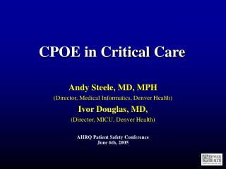
Normal Ultrasound Pattern The pleural line (white arrow) is a roughly horizontal hyperechoic line 0.5 cm below the upper and lower ribs identified by acoustic shadow (R). A single vertical artifact arising from the pleural line and spreading up to the edge of the screen (comet-tails, indicated by asterisk) can be seen in dependant regions in normally aerated lungs
Normal Ultrasound Pattern 'lung sliding' associated with artifactual horizontal A-lines
Ultrasound Aspects of Alveolar-Interstitial Syndrome B-lines 7 mm apart or spaced comet-tail artifacts. The pleural line (white arrow) and the ribs (R) with their acoustic shadow. B-lines arising from the pleural line and spreading up to the edge of the screen correspond to thickened interlobular septa .
Ultrasound Aspects of Alveolar-Interstitial Syndrome B-lines 7 mm apart or spaced comet-tail artifacts. These artifacts correspond to thickened interlobular septa .
Ultrasound Aspects of Alveolar-Interstitial Syndrome B-lines 3 mm or less apart. The pleural line (white arrow) and the rib (R) with their acoustic shadow. Contiguous comet-tails arising from the pleural line and spreading up to the edge of screen correspond to ground-glass areas on chest CT scan.
Ultrasound Aspects of Alveolar-Interstitial Syndrome B-lines 3 mm or less apart. The pleural line (white arrow) and the rib (R) with their acoustic shadow. Contiguous comet-tails arising from the pleural line and spreading up to the edge of screen are present. These artefacts correspond to ground-glass areas on chest CT scan.
Ultrasound Aspect of a Lung Consolidation and Pleural Effusion Transversal view of consolidated left lower lobe; lung consolidation is seen as a tissular structure (C). In this consolidation, hyperechoic punctiform images (indicated by asterisk) can be seen; these correspond to air bronchograms (air-filled bronchi). Pleural effusion is anechoic (Pl). .
Ultrasound Aspect of Lung Consolidation and Pleural Effusion Cephalocaudal view of consolidated left lower lobe: lung consolidation with air bronchograms. Ao, descending aorta; D, diaphragm; Pl, pleural effusion..
Ultrasound Aspect of Lung Consolidation and Pleural Effusion Cephalocaudal view of consolidated left lower lobe: lung consolidation with air bronchograms.
Consolidated Left Lower Lobe with a Peripheral Abscess. The abscess (A) appears as rounded hypoechoic lesions inside a lung consolidation (C). Ao, descending aorta; D, diaphragm; Pl, pleural effusion. .
Consolidated Left Lower Lobe with a Peripheral Abscess. The abscess (A) appears as rounded hypoechoic lesions inside a lung consolidation (C). Ao, descending aorta; D, diaphragm; Pl, pleural effusion. .
Consolidated lung 'floating' in a Massive Pleural Effusion The pleural effusion (Pl) is abundant enough to be compressive and the lung (C) is seen consolidated and floating in the pleural effusion.
Consolidated Lung and Adjacent Pleural Effusion with Pleural Adherences. The pleural effusion (Pl) is abundant and the lung is seen consolidated and floating (C) in the pleural effusion with pleural adherences. (A)
Pneumothorax with “Lung Point” (a) Normal lung and (b) pneumothorax patterns using time-motion mode lung ultrasound. In time motion mode, one must first locate the pleural line (white arrow) and, above it, the motionless parietal structures. Below the pleural line, lung sliding appears as a homogenous granular pattern (a). In the case of pneumothorax and absent lung sliding, horizontal lines only are visualised (b). In a patient examined in the supine position with partial pneumothorax, normal lung sliding and absence of lung sliding may coexist in lateral regions of the chest wall. In this boundary region, called the 'lung point' (P), lung sliding appears (granular pattern) and disappears (strictly horizontal lines) with inspiration when using the time-motion mode
US Sensitivity and Specificity for Pneumothorax Lichtenstein, DA; Meziere, G; Lascols, N; Biderman, P; Courret, JP; Gepner, A; Goldstein, I; Tenoudji-Cohen, M. Ultrasound diagnosis of occult pneumothorax. Crit Care Med. 2005;33:1231–1238.
The problem... • Complications • Pneumothorax • Hemothorax • Arterial puncture • Hematoma formation • Neck, groin, mediastinum • Failure to obtain access
Complications McGee DC, Gould MK. Preventing Complications of Central Venous Catheterization. NEJM 2003;348:1123-33
Ultrasound Guided Vascular Access • Agency for Healthcare Research and Quality • Making Health Care Safer: A Critical Analysis of Patient Safety Practices • “Use of real-time ultrasound guidance during central line insertion to prevent complications” http://www.ahcpr.gov/clinic/ptsafety/chap21.htm
Ultrasound Guided Vascular Access “In hospitals where US equipment is available and physicians have adequate training, the use of US guidance should be routinely considered for cases in which IJ venous catheterization will be attempted” McGee DC, Gould MK. Preventing Complications of Central Venous Catheterization. NEJM 2003;348:1123-33
Adult IJ – US vs Landmark Hind DH, Calvert N, Davidson A, et al. BMJ 2003
Adult IJ • Denys et al “randomized” patients to IJ- US guided=928, Landmark=302 • Overall success 100% vs 88.1% • First attempt success 78% vs 38% • Skin to vein time 9.8 (2-68) vs 44.5 (2-1000) sec • Carotid puncture 1.7% vs 8.3% Denys BG, Uretsky BF, Reddy PS. Ultrasound-assisted cannulation of the internal jugular vein – a prospective comparison to the external landmark-guided technique. Circulation 1993;87:1557-62
Vein Thinner walls Compressible Non-pulsatile (color flow) Vein Versus Artery • Artery • Thicker walls • Non-compressible • Pulsatile • (color flow)
Ultrasound Access Techniques • “Static” • mapping technique • no sterile technique required for US • “Dynamic” • views needle entering vein • freehand • needle guide • requires sterile technique
Static Technique • Position patient as you will for procedure • Look at vessels and confirm landmark-predicted anatomy • Mark location, note depths and angles • Remove ultrasound, prep patient without moving • Vein cannulated as usual
Dynamic Technique • Place gel in palm of sterile glove • Place vascular probe in palm, avoid trapped air bubbles, and wrap free fingers out of way • Sterile KY jelly for glove-skin interface
Dynamic technique • Center vessel in center of screen • Center of probe overlies center of vessel and serves as landmark for needle • Needle creates bright echo with ringdown artifact • Advance needle to vein, which is deformed by needle pressure and then recoils to original position as vein is cannulated
Internal Jugular Medial Lateral IJ Carotid
Internal jugular Medial Lateral
Internal Jugular Medial Lateral
Goals of FAST • Rapid detection of: • Hemoperitoneum • Hemopericardium • Hemothorax • Advanced/expanded • rapid detection of pneumothorax • identification of solid organ injury
Standard Views RUQ Morison’s View LUQ View Pelvic View
RUQ Diaphragm Liver Head Foot Lower pole kidney Upper pole kidney
Free Fluid in Morison’s Pouch Very rough rule of thumb: 0.5 cm fluid stripe = 500 cc, 1 cm fluid stripe = 1 L
False + RUQ view IHepatic Vein IVC