Download
1 / 27
270 likes | 766 Views
Explore the etiology, signs, symptoms, and treatment of invasive cancer of the vulva, including risk factors, histologic types, and staging information. Stay informed on this critical aspect of women's health.

E N D
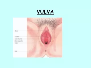
INVASIVE CANCER of THE VULVA RuksetAttar, MD, PhD ObstetricsandGynecology Department
incidence has been increasing: 8 % • Usu at age 65-75 years • Cases should be classified as carcinoma of the vulva when the primary site of the growth is in the vulva. • A carcinoma of the vulva that has extended to the vagina should be considered as a carcinoma of the vulva. • Malignant melanoma should be reported separately.
Risk factors DM Obesity HT-40% and atherosklerosis Nulliparity Premature menapause Sy Abnormal PAP smear test Age
Working at a laundry or dry cleaning Vulvardysthrophies HPV infection CIN Cigarette smoking Immunsupression Genital wart VIN- 30% P53, PTEN, Rb gen mutation
Incidence of vulvar neoplasms by histologic type TumortypePercent Epidermoid 86.2 Melanoma 4.8 Sarcoma 2.2 Basalcell 1.4 Bartholingland 1.2 squamous 0.4 adenocarcinoma 0.6 Adenocarcinoma 0.6 Undifferentiated 3.9
ETIOLOGY • HumanpapillomavirusType 16,18,31 • Coal tar chemicals • Arsenicdermatitis-in laundrydetergents • Vulvarirritation • Anychronicvulvitis • Herpeticvulvitis ?
In terms of etiology, pathogenesis, and clinical presentation, vulvarsquamous cell carcinomas divided into two general groups • The first group • in youngerpatients • associated with cancer-related HPVandsmoking • multicentric, • frequently coexists with or is preceded by a classic Vulvar IntraepithelialNeoplasia (VIN). • A variety of chromosome abnormalities are linked to invasive vulvar cancer, some of whichmay be specific for HPV-positive tumours.
The second group • Inelderlyparients • associated with squamous cell hyperplasia and lichen sclerosis. • The etiology of this group of carcinomas is unclear, infrequently associated with HPV: • genetic alterations arise in lichen sclerosus or hyperplasia, leading directly to invasion, or • atypia develops within hyperplasia or lichen sclerosis (differentiated VIN). • These tumours areassociated with mutations in p53 • significantly worse prognosis than HPV-positive tumours
SIGNS AND SYMPTOMS OF VULVAR CANCER Signs and symptoms Percent Pruritis 45.0 Mass 45.0 Pain 23.0 Bleeding 14.0 Ulceration 14.0 Dysuria 10.0 Discharge 8.0 Groin mass 2.5
Indications for excisional biopsy of vulvar nevi • Change in surfacearea of nevus • Change in elevation of a lesion – raised,thickened,ornodular • Change in color – especiallybrowntoblack • Change in surface – smoothtoscalyorulcerated • Change in sensation – itchingortingling
FIGO Staging of Vulva CA • Stage I Tumor confined to the vulva • IA Lesions ≤2 cm in size, confined to the vulva or perineum and with stromal invasion ≤1.0 mm, no nodal metastasis • IB Lesions ›2 cm in size or with stromal invasion › 1.0 mm, confined to the vulva or perineum, with negative nodes • Stage II Tumor of any size with extension to adjacent perineal structures (1/3 lower urethra, 1/3 lower vagina, anus) with negative nodes
Stage III Tumor of any size with or without extension to adjacent perineal structures (1/3 lower urethra, 1/3 lower vagina, anus) with positive inguino-femoral lymph nodes • IIIA • (i) With 1 lymph node metastasis (≥5 mm), o • (ii) 1–2 lymph node metastasis(es) (‹5 mm) • IIIB • (i) With 2 or more lymph node metastases (≥5 mm), or • (ii) 3 or more lymph node metastases (‹5 mm) • IIIC With positive nodes with extracapsular spread
Stage IV Tumor invades other regional (2/3 upper urethra, 2/3 upper vagina), or distant structures • IVA Tumor invades any of the following: • (i) upper urethral and/or vaginal mucosa, bladder mucosa, rectal mucosa, or fixed to pelvic bone, or • (ii) fixed or ulcerated inguino-femoral lymph nodes • IVB Any distant metastasis including pelvic lymph nodes • The depth of invasion is defined as the measurement of the tumor from the epithelial stromal junction of the adjacent most superficial dermal papilla to the deepest point of invasion.
Routesof Spread • Direct extension • vagina,urethra,anus • Lymphatic embolization • regional lymph nodes • Hematogenous spread • distant sites,lung,liver,bone
Risk of metastatic spread size of tumour depth of invasion involvement of lymphatic vessels. • most commonly involved nodes: inguinal, femoral, pelvic, iliac, and periaortic lymph nodes. • hematogenous dissemination involves the lungs, liver, and other internal organs
Therapy • Staging and treatment for stage I and II vulvar cancer is surgical • The primary treatment is complete surgical removal • Stage Ia: wide local excision (2 cm margin) • Stage Ib: radical local excision (3-4 cm margin) with lenfadenecytomy with adjuvant RT • Stage ≥II • pelvic egzenteration • Radical vulvectomy with lenfadenecytomy with or without preopchemoRT (cisplatin-5FU) or postop RT
INVASIVE CANCER of THE VAGINA RuksetAttar, MD, PhD ObstetricsandGynecology Department
Age 60 years More than 50% in posterior and 1/3 upper vaginal wall May be due to chronic irritation Symptoms as in cervix ca Vaginal esp bloody discharge Urinary symptoms Pelvic pain
85% are epidermoid cancers, and the remainder, in decreasing order of frequency, are adenocarcinomas, sarcomas, and melanomas. A tumor should not be considered a primary vaginal cancer unless the cervix is uninvolved or only minimally involved by a tumor obviously arising in the vagina. Any malignancy involving both cervix and vagina that is histologically compatible with the origin in either organ is classified as cervical cancer.
Secondary carcinoma of the vagina is seen more frequently than primary vaginal cancers. Secondary, or metastatic, tumors may arise from cervical, endometrial, or ovarian cancer, gestational trophoblastic disease, colorectal cancer, or urogenital or vulvar cancer. Extension of cervical cancer to the vagina is probably the most common malignancy involving the vagina.
Risk Factors HPV Pesser Vaginal prolapsus Chronic leukorrhea sy early hysterectomy prior radiation are possible no specific etiologic agent has been identified
Local invasion • Lymphatic spread • Upper vaginal wall- obturatory, iliac and hypogastric and paraaortic- as in cervix • Lower vagina-inguinal, pelvic-as in vulva • Hematogeneus spread • Lung, liver, bone
FIGO staging of carcinomaof the vagina • Stage 0 Carcinoma in situ, intraepithelial carcinoma. • Stage I limited to the vaginal mucosa • Stage II involved the subvaginal tissue • A but not extended to real paracolpium • B involved real paracolpium but not extended to the pelvic wall • Stage III extended to the pelvic wall or symphsis involvement • Stage IV extended beyond the true pelvis or has involved the mucosa of the bladder or rectum. • A Spread of the growth to adjacent organs- the mucosa of the bladder or rectum • B Spread to distant organs.
Diagnosis is difficult Abnormal PAP smear without lesion in vulva or cervix Colposcopy Biopsy Clinical Staging
Therapy • Upper vaginal wall ca • Radical hysterectomy with vagenectomy • RT • Surgery with RT • Lower vaginal wall ca • Radical vulvectomy with vagenectomy • RT • Surgery with RT • Stage IVa- egzenteration