Download
1 / 27
270 likes | 311 Views
Explore the discovery of x-ray, electromagnetic radiation origins, and different imaging modalities like CT, MRI, and ultrasound. Understand the principles behind each modality in medical imaging.

E N D
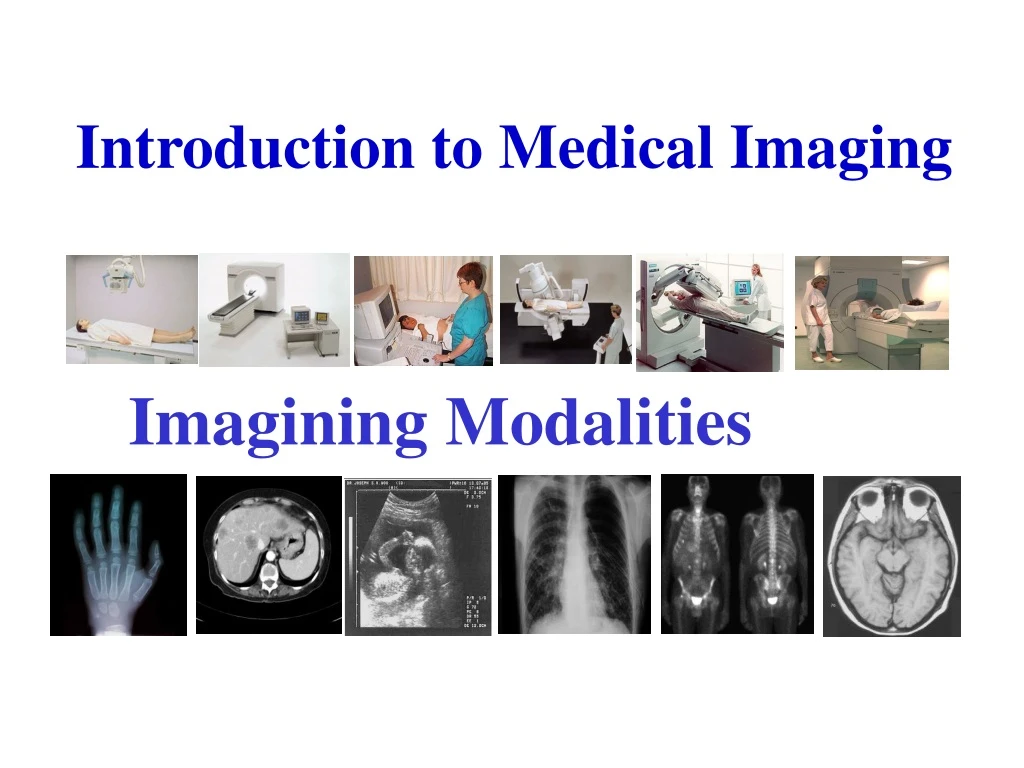
Introduction to Medical Imaging Imagining Modalities
Learning Objectives By the end of this Lecture the student will be able to: • Discus the discovery of x-ray • the origin of electromagnetic radiation • Identify the different modalities • X-ray ( radiography/Fluoroscopy) • Computerized Tomography (CT) • Nuclear Medicine, PET and SPECT • Ultrasound (U/S) • Magnetic Resonance Imaging (MRI) • 3. Understand the basic principles for each imaging modality
References • Text book of radiographic positioning and related anatomy; by Kenneth • L.Bontrager, 5th edition • Useful Websites • http://www.e-radiography.net/ http://faculty.ksu.edu.sa/74344/default.aspx
Who discovered the x-ray? • On November 8th, 1895, German scientist Wilhelm Roentgen was • conducting experiments in his laboratory on the effects of cathode rays. • Specifically he was observing the effect of passing an electrical discharge • through gases at a low pressure. While doing so, Roentgen noticed when • passing current through the cathode ray, rays were given off that passed • through materials such as wood, paper and aluminum. • The Roentgen conclusion from his experiments is (unknown type of radiation) For that he name it X-RAYS X meaning unknown electromagnetic radiation of short wavelength produced when high-speed electrons strike a solid target
Imagining Modalities • Radiography (X-Ray) • Fluoroscopy (guided procedures) • Diagnostic / Interventional Transmission imaging • Computed Tomography (CT) • Ultrasound (US) • Gray-Scale /Color Doppler Reflection imaging • Magnetic Resonance Imaging (MRI) • Nuclear Medicine (Gamma/ PET/SPECT ) Emission imaging
High Electrical Potential Electrons - + Radiation Penetrate the Sample Exposure Recording Device Conventional X-ray Imaging. X-ray Production. Electrons from cathode filament are accelerated towards and impact the rotating anode. Rapid deceleration produces heat (~ 98%) and x-rays (~2%)
Over couch X-ray Tube and Table High Tension Cables X-ray Tube housing Controls Light Beam Diaphragm Table, and cassette holder
Conventional X-ray Image of a Hand Normal Arthritic
Fluoroscopy Fluoroscopy is a study of moving body structures - similar to an x-ray "movie." A continuous x-ray beam is passed through the body part being examined, and is transmitted to a TV-like monitor so that the body part and its motion can be seen in detail.
Fluoroscopy TV CAMERA OR CCD ARRAY (for digital screening) IMAGE INTENSIFIER X-RAY TUBE Allows dynamic imaging of blood vessels (angiography) and ‘interventional’ procedures CONTROLS
Computerized Tomography (CT) CT imaging combines special x-ray equipment with sophisticated computers to produce multiple images of the inside of the body. These cross-sectional images of the area being studied can then be examined on a computer monitor or printed. Brain Axial Image CT Scanner
Computed Tomography (CT) X-ray tube Rotation gives multiple projections Thin fan beam of x-rays Patient (stationary) Array of detectors (rare-earth doped ceramics with photodiodes)
Radioisotope Imaging Nuclear medicine(NM) IN Radioisotope imaging the formation of images provides information about the function of various organs in the body, using internally administered radioisotopes as a radiation source. The technique is widely used in medicine to locate tumors or cancers and to examine the flow patterns of body fluids. Gamma camera head
Radioisotope Imaging Positron Emission Tomography (PET) Positron emission tomography (PET) is a nuclear medicine imaging technique which produces a three-dimensional images of functional processes in the body. The system detects pairs of gamma rays emitted indirectly by a positron-emitting radionuclide (tracer), which is introduced into the body on a biologically active molecule. Images of tracer concentration in 3-dimensional space within the body are then reconstructed by computer analysis. In modern scanners, this reconstruction is often accomplished with the aid of a CT X-ray scan performed on the patient during the same session, in the same machine.
“Dead” areas of brain No glucose metabolism Radioisotope Imaging Single Photon Emission Computed Tomography (SPECT) Is a nuclear medicine tomographic imaging technique using gamma rays. It is very similar to conventional nuclear medicine planar imaging using a gamma camera. However, it is able to provide true 3D information. This information is typically presented as cross-sectional slices through the patient, but can be freely reformatted or manipulated as required. Human Brain - Stroke
Ultrasound Imaging (US) Ultrasound imaging is a common diagnostic medical procedure that uses high-frequency sound waves to produce images sonograms) of organs, tissues, or blood flow inside the body. The procedure involves using a transducer, which sends a stream of high-frequency sound waves into the body and detects their echoes as they bounce off internal structures. The sound waves are then converted to electric impulses, which are processed to form an image displayed on a computer monitor. It is from these images that videos and portraits are made. Ultrasound Transducer
Magnetic Resonance Imaging (MRI) MR imaging uses a powerful magnetic field, radio frequency pulses and a computer to produce detailed pictures of organs, soft tissues, and all other internal body structures. The images can then be examined on a computer monitor, printed or copied to CD. MRI does not use ionizing radiation (x-rays). Big superconducting magnet (~ 1.5 tesla). Gradient coils. Radiofrequency coils.
Magnetic Resonance Imaging (MRI) Axial Brain Images Proton density weighted T2-weighted T1-weighted
Safety Radiation Type Modality Comments } X-ray imaging Radioisotope scanning Ultrasound Imaging MRI Biological effect , need protection against unnecessary doses Ionising Radiation } Non-ionising Radiation Less harmful effects. Better for the foetus.