DENTIN
DENTIN. Provides the bulk and general form of tooth. Determines the shape of the crown. Physically & chemically the dentin closely resembles the bone.

DENTIN
E N D
Presentation Transcript
DENTIN • Provides the bulk and general form of tooth. • Determines the shape of the crown. • Physically & chemically the dentin closely resembles the bone. • The main morphologic difference between bone & dentin is that some of the osteoblasts that form bone marrow enclosed within its matrix substance as osteocytes, whereas the dentin contains only the processes of the cells that form it. • Both are considered vital tissue because they contain because they contain living protoplasm.
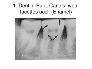
PHYSICAL PROPERTIES • It is light yellowish in color, becoming darker with age. • It is elastic and subject to slight deformation. • Harder than bone but softer than enamel. • Lower content of mineral salts in dentin renders it more radiolucent than enamel.
CHEMICAL PROPERTIES • Consists of 35% organic matter and water & 65% inorganic material. • The organic substance consists of collagenous fibrils and a ground substance of mucopolysaccharides (proteoglycans and glycos aminoglycans). • The inorganic component consists of hydroxyapetite as in bone, cementum & enamel. • Organic constituents can be removed from the mineral by incineration or organic chelation.
STRUCTURE • The bodies of the odontoblasts are arranged in a layer on the pulpal surface of the dentin, and only their cytoplasmic processes are included in the tubules in the mineralized matrix. • Each cell gives rise to one process, which traverses the predentin & calcified dentin within one tubule. • Terminates in a branching network at the junction with enamel or cementum. • Tubules are found throughout normal dentin & are therefore characteristic of it.
DENTINAL TUBULES • The course follows a gentle curve in the crown, less so in the root, where it resembles S in shape. • Starting at right angles from the pulpal surface, the first convexity of this doubly curved course is directed toward the apex of the tooth. • These tubules end perpendicular to the dentinoenamel and dentinocementum junctions. • Near the root tip & along the incisal edges and cusps the tubules are almost straight.
The ratio between the outer and inner surfaces of dentin is about 5:1. • The ratio between the numbers of tubules per unit area on the pulpal and outer surfaces of dentin is about 4:1. • There are more tubules per unit area in the crown than in the root. • The dentinal tubules have lateral branches throughout dentin, which are termed canaliculi or microtubules. • A few dentinal tubules extend through the dentinoenamel junction into the enamel. These are termed enamel spindles.
PERITUBULAR DENTIN • The dentin that immediately surrounds the dentinal tubules. • It is more highly mineralized than intertubular dentin. • It is twice as thick in outer dentin (approx. 0.75um) than in inner dentin (0.4um). • By its growth, it constricts the dentinal tubules to a diameter of 1um near the dentinoenamel junction. • Organic matrix is lost along with mineral after decalcification. • The calcified tubule wall has an inner organic lining termed the lamina limitans, high in glucosaminoglycans (GAG).
INTERTUBULAR DENTIN • Forms the main body of dentin. • It is located between the dentinal tubules or, more specifically, between the zones of peritubular dentin. • Its organic matrix is retained after decalcification. • About one-half of its volume is organic matrix, specifically collagen fibers. The fibrils range from 0.5 to 0.2um in diameter and exhibit crossbanding at 64um intervals, which is typical for collagen.
PREDENTIN • Is located adjacent to the pulp tissue. • Is 2 to 6 um wide, depending on the activity of the odontoblast. • It is the first formed dentin and is not mineralized. • As the collagen fibers undergo mineralization at the predentin- dentin front, the predentin then becomes dentin and a new layer of predentin forms circumpulpally.
ODONTOBLAST PROCESS • They are the cytoplasmic extensions of the odontoblasts. • The odontoblasts reside in the peripheral pulp at the pulp- predentin border and their processes extend into the dentinal tubules. • The processes are largest in diameter near the pulp and taper further into dentin. • The odontoblast cell bodies are approximately 7um in diameter and 40um in length.
RELATIONSHIP BETWEEN ODONTOBLASTIC PROCESS AND DENTINAL TUBULE
PRIMARY DENTIN • Mantle dentin is the first formed dentin in the crown underlying the dentinoenamel junction. • It is the outer or most peripheral part of the primary dentin & is about 20um thick. • The fibrils found in this zone are perpendicular to the dentinoenamel junction. • Circumpulpal dentin forms the remaining primary dentin or bulk of the tooth. • Represents all of the dentin formed prior to root completion. • The fibrils are much smaller in diameter & are more closely packed together. • Slightly more mineral content than mantle dentin.
SECONDARY DENTIN • A narrow band of dentin bordering the pulp and representing the dentin formed after root completion. • Contains fewer tubules than primary dentin. • There is usually a bend in the tubules where primary and secondary dentin interface.
INCREMENTAL LINES • The incremental lines (von ebner), or imbrication lines, appear as fine lines or striations in dentin. • They run at right angles to the dentinal tubules. • These lines reflect the daily rhythmic, recurrent deposition of dentin matrix as well as hesitation in the daily formative process. • The course of the lines indicates the growth pattern of the dentin. • Some of the incremental lines are accentuated because of disturbances in the matrix and mineralization process. Such lines are known as contour lines of owen.
These lines represent hypocalcified bands. • In the deciduous teeth and in the first permanent molars, the prenatal and postnatal dentin is separated by an accentuated contour line. This is termed the neonatal line. • This line reflects the abrupt change in environment that occurs at birth. • The dentin matrix formed prior to birth is usually of better quality than that formed after birth.
INTERGLOBULAR DENTIN • Sometimes mineralization of dentin begins in small globular areas that fail to fuse into a homogenous mass. This results in zones of hypomineralization between the globules. These zones are called interglobular dentin. • Forms in crowns of teeth in the circumpulpal dentin just below the mantle dentin. • Follows an incremental pattern. • The dentinal tubules pass uninterruptedly, thus demonstrating a defect of mineralization & not of matrix formation.
GRANULAR LAYER • There is a zone adjacent to the cementum that appears granular. This is known as Tomes’ granular layer. • Slightly increases in amount from the cementoenamel junction to the root apex. • Caused by coalescing and looping of the terminal portions of the dentinal tubules. • The odontoblast initially interacts with ameloblasts or root sheath cells through the basal lamina.
THEORIES OF PAIN TRANSMISSION THROUGH DENTIN • Direct conduction theory in which stimuli directly effect the nerve endings in the tubules. • Transduction theory in which the membrane of the odontoblast process conducts an impulse to the nerve endings in the predentin, odontoblast zone, and pulp. • Fluid or hydrodynamic theory in which stimuli cause an inward or outward movement of fluid in the tubule, which in turn produces movement of the odontoblast and its processes.
AGE AND FUNCTIONAL CHANGES VITALITY OF DENTIN • Odontoblasts and its processes are an integral part of dentin. • Reacts to physiologic and pathologic stimuli. REPARATIVE DENTIN • Also known as tertiary or response dentin. • If by extensive abrasion, erosion, caries, or operative procedures the odontoblast processes are exposed or cut, the odontoblasts die or, if they live, deposit reparative dentin. • Origin of the new odontoblast is from undifferentiated perivascular cell. • Has fewer & more twisted tubules than normal dentin. • Sometimes, a combination of osteodentin & tubular dentin is seen. • Odontoblasts lay down 1.4 um/day.
An accentuated calcio-traumatic band under the cavity represents an acute arrest inodontoblastic activity as a result of the operative procedure
The following notations are used for the assessment of the acute calcio-traumatic effect. • O-Normal. Calcio-traumatic effect absent or no difference between the calcio-traumatic effect under the cavity preparation and the control areas or control tooth. • 1-Faint accentuation of the calcio-traumatic line under the cavity as compared to control areas; reflects a slight injury to the odontoblasts. • 2-Definite accentuation of the calcio-traumatic line under the cavity; reflects a definite acute injury to the odontoblasts.
CLASSIFICATION OF DENTIN ON THE BASIS OF LOCATION, PATTERN OF MINERALIZATION AND DEVELOPMENTAL PATTERN.
DEAD TRACTS • The odontoblast processes disintegrate, & the empty tubules are filled with air. • Appear black in transmitted light & white in reflected light. • Often observed in the area of narrow pulpal horns because of crowding of odontoblasts. • Demonstrate decreased sensitivity. • Appear to a greater extent in older teeth. • Probably the initial step in the formation of sclerotic dentin.
SCLEROTIC OR TRANSPARENT DENTIN • Collagen fibers and apatite crystals begin appearing in the dentinal tubules. • Apatite crystals are initially only sporadic in a dentinal tubule but gradually fill it with a fine meshwork of crystals. • Gradually, the tubule lumen is obliterated with mineral, which appears very much like the peritubular dentin. • The refractive indices of dentin in which the tubules are occluded are equalized, and such areas become transparent. • Found specially in roots. • Transparent or light in transmitted and dark in reflected light.
Clinical considerations • The rapid penetration & spread of caries in the dentin is the result of the tubule system in the dentin. • The dentinal tubules form a passage for invading bacteria that may thus reach the pulp through a thick dentinal layer. • Air driven cutting instruments cause dislodgement of the odontoblasts from the periphery of the pulp & their aspiration within the dentinal tubule. • Sensitivity of dentin. • Effect of the smear layer.
Affected & infected dentin • Infected dentine is the outer layer and is softened and contaminated with bacteria. It is irreversibly denatured and not remineralized • Affected dentine has a demineralized phase, but not yet invaded by bacteria. It can be remineralized. • In clinical restorative treatment of dentine during cavity preparation it is infected dentine which is completely removed. The affected dentine, which may be remineralized after the completion of restorative treatment, is not removed and is preserved.
Caries-affected dentin interfaces showed a much thicker hybrid layer than was seen with non-carious dentin. • Presumably, the thicker hybrid layer in caries-affected dentin may be due to the fact that caries-affected dentin is partially demineralized and more porous than non-carious dentin. • Allow deeper penetration of the acid etchant, leading to a deeper demineralization with diffused monomer.