Download
1 / 32
330 likes | 621 Views
Kainate Receptors & Synaptic Plasticity. Sorleen Trevino and Eamon Quick 12/4/06. GluR5 subunit structure, www.dfuni.dk. Kainate & Synaptic Plasticity. Three types of fast glutamate receptors: NMDA AMPA Kainate

E N D
Kainate Receptors & Synaptic Plasticity Sorleen Trevino and Eamon Quick 12/4/06 GluR5 subunit structure, www.dfuni.dk
Kainate & Synaptic Plasticity • Three types of fast glutamate receptors: • NMDA • AMPA • Kainate • Interest in kainate has been ever-increasing, and has roles in synaptic transmission and synaptic plasticity. • While NMDA-dependent plasticity has often been the de facto model of synaptic facilitation, kainate has been found to not only been involved in the phenomenon, but to have unique features to contribute to synaptic plasticity. • Some novel features of kainate-dependent synaptic plasticity include: • Heterosynpatic facilitation** • Presynaptic bi-directional modulation • Low-frequency enhancement of synaptic strength** ** These features are somewhat controversial…
Pre or Postsynaptic Expression? • Kullmann and Siegelbaum explain: • Both actually… • And some may disagree on specifics, • Bias experiments… • LTP and LTD is often dependent on NMDA receptor activation. • “strong” brief high-frequency tetanic stimulation (HFS) induces LTP • “weaker” prolonged low-frequency stimulation (LFS) induces LTD.
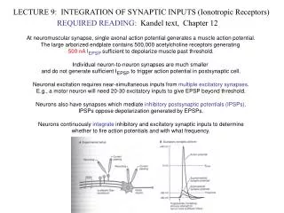
EC stimulation activates excitatory afferents from cortical structures, including the lateral entorhinal and temporal cortices, that course through the EC and synapse in the BSA. LFS in the BLA slice can induce potentiation or depression; it shows an experience-dependent switch (metaplasticity), and is not dependent on NMDARs, but on group II metabotropic glutamate receptors. Modification of activity-dependent LTP adds flexibility could play a role emotional memory and regulation of epileptogenesis.
LFS and HFS induces synaptic plasticity in the BSA • 1 Hz for 15 min; LFS induced an enduring enhancement; maintained for more than 30 mins • Brief HFS; 100 Hz, 1 sec; induced APV-sensitive short-term potentiation. • LFS after HFS short-term potentiation (HFS/LFS), causes enhancement initially and is then reversed to persistent amplitude depression. • Stimulus-dependent long-lasting synaptic depression in the amygdala. LFS HFS LFS HFS HFS Li 1998
De-potentiation of enhancement by LFS only after HFS • The first presentation of LFS produced an enduring facilitation • A second LFS train failed to induce depression, nor did the second train produced further facilitation. • LFS induced an enduring facilitation, subsequent HFS caused further transient potentiation, but a second LFS train applied after HFS resulted in de-potentiation. • There was always a brief period of enhancement during LFS before the onset of de-potentiation. • The direction of the long-term change in synaptic efficacy induced by LFS was found to be dependent on the history of previous activation. LFS HFS LFS
Mechanism of LFS is NMDAR independent • LFS-induced enduring facilitation is NMDAR independent • 100 uM APV does not alter the amplitude of the synaptic response • Brief HFS (100 Hz for 1 sec) induces NMDAR-dependent short term enhancement that is reduced by APV; it shows that short-term potentiation in the amygdala is dependent on NMDAR activation. • HFS/LFS-induced synaptic depression is NMDAR independent • APV nor MK-801 do not affect the HFS/LFS-induced depression LFS LFS HFS LFS HFS
EGLU prevents HFS-dependent switch from facilitation to depression • Drug has no antagonist effects on postsynaptic metabotropic and ionotropic glutamate receptors • Application of presynaptic (group II) metabotropic glutamate receptor antagonist EGLU prevents HFS/LFS induced depression • It did not alter LFS-induced enduring facilitation, in contrast to the synaptic depression ordinarily observed • EGLU block can be attributed to an action on the HFS priming mechanism since the block seems to affect only the depression LFS HFS LFS HFS LFS HFS
Differences in Amygdala LTP: • In the basolateral amygdala, we find that the history of synaptic activity rather than the frequency, determines the direction of the enduring modification in synaptic strength. • Effects of activity on plasticity is referred to as “metaplasticity,” a higher-order form of synaptic plasticity (Abraham and Tate, 1997). • Important differences from conventional LTP (like that of hippocampal CA1): • LFS-induced enhancement develops slowly in response to prolonged stimulation. • LFS-induced enhancement occurs monotonically, one step. • In contrast to hippocampal LTP, amygdala LFS-induced enhancement is additive with STP (short-term). • LFS-induced enhancement is not dependent on NMDA receptors; the switch from facilitation to depression could be dependent on class II presynaptic metabotropic glutamate receptors. • Synaptic depression in the amygdala is only induced when LFS occurs previous to HFS; mechanism remains to be determined. • Similarities with LTP that is not NMDAR-dependent, like in CA3. • Explain this phenomenon: presynaptic metabotropic glutamate receptors do feedback inhibition of release, resulting in depression, which is why EGLU inhibits HFS/LFS-induced depression.
New antagonist introduced, LY382884. • selectively antagonizes GluR5 containing kainate receptors in a concentration dependent manner • doesn’t affect AMPAR or NMDAR; 10uM is the max concentration before AMPAR transmission is affected • CA3 neurons possess postsynaptic kainate Rs and are high in density in CA3 area • In situ hybridization histochemistry shows dense expression of GluR5 mRNA in the amygdala, especially basolateral cortex, and adjacent piriform cortex. • GluR5 expression is higher in the amygdala than in the hippocampus, opposite for GluR6 and KA-2 mRNA. • Results confirmed by RT-PCR of GluR5 mRNA extracted from microdissections of slices • Explore kainate R importance in LTP induction at mossy fibre synapses • Found that LY382884 has no effect on NMDAR- dependent LTP, but prevents mossy fibre LTP, which is NMDAR independent.
LY382884 blocks GluR5-KAR and MF LTP • ATPA is a selective GluR5 kainate receptor ligand. • LY382884 antagonizes the depression of AMPAR-mediated EPSPs induced by ATPA. • Mossy fibre LTP was completely, but reversibly, prevented by LY382884. • CNQX and kynurenate at a concentration that also antagonized GluR5 kianate current also blocks mossy fibre LTP
Mossy fibre LTP involves activation of the cAMP-PKA pathway which is induced by adenylyl cyclase stimulation via forskolin. • LY382884 has no effect on forskolin-induced mossy fibre LTP. • Kynurenate inhibits AMPAR-mediated transmission, but upon washout does not prevent induction of mossy fibre LTP. • GYKI53655, a more selective AMPAR antagonist shows the same results.
These three compounds antagonize kainate receptors containing GluR5 and MF LTP in the ranking order of potency: LY382884 = CNQX >> kynurenate. • Kainate receptor component of excitatory transmission at mossy fibre synapses is antagonized by GluR5 antagonists and absent in GluR6 knockout mice, GluR6 also crucial. • As for the question of whether mossy fibre LTP is induced pre- or post-synaptically, it is hard to say because GluR5-containing kainate receptors are located at both sites. • Show that kainate receptors are involved in LTP induction at a synapse where NMDA receptors don’t have a role.
LFS-induced facilitation • Induction of enduring enhancement in BSA neurons by LFS of EC requires activation of GluR5 kainate-type ionotropic glutamate receptors acting through a Ca2+ dependent mechanism. • LFS of EC facilitates BSA EPSPs, as shown by Li 1998. LFS
LFS-induce facilitation eliminated by GluR5 kainate R antagonist - as seen in Bortolotto 1999 • Component of depolarization is mediated by GluR5 kainate receptors, which is resistant to NMDAR and AMPAR antagonists and is blocked by decahydroisoquinoline/LY377770, which is like LY382884, and selectively blocks GluR5 kainate receptors. • In the presence of APV, additional perfusion with GYKI 53655 (AMPAR antagonist), bicuculline and SCH 50911 (GABAa and GABAb R antagonists) reduces the response amplitude to its kainite receptor component, which is eliminated by 20 uM LY377770. • GluR5 kainate receptors are required for the induction of low-frequency stimulation-induced synaptic facilitation in the BSA. LFS LFS
Plasticity mimicked by GluR5 kainate receptor agonist, ATPA • ATPA-enhancement was prolonged as it lasted longer than 120 min and did not decrement. • It also seemed to have several components, meaning possible recruitment of excitatory synaptic potentials. Although the initial component is monosynaptic. • Also, after perfusion there was an greater increase in slope vs peak amplitude. • ATPA initially causes depression but recovers and increases even more after perfusion. • Inclusion of the LY377770 eliminates the delayed enhancement, but the initial ATPA-induced depression was not blocked meaning this depression is not mediated by GluR5 kainate receptors. • ATPA is also very similar to AMPA, and can activate AMPARs, but this facilitation is not mediated by AMPARs because AMPA application causes depression rather than facilitation.
Hetrosynapticism • This GluR5 kainate R-mediated facilitation develops slowly and also enhances adjacent unstimulated/inactive afferents from the BA.
Role of Ca++ in heterosynaptic facilitation • GluR5 kainate receptor pre-mRNA that undergo post-transcriptional nuclear editing, have their arginine (R) substituted for a glutamine (Q) at residue 636 within the second hydrophobic (MII) domain. • GluR5 kainate receptors with unedited subunits are more Ca2+ permeable. • RT-PCR of the BLA that is further subjected to and cleaved by the restriction endonuclease BbVI to quantify regional GluR5 mRNA. • The heterosynaptic enhancement of EC- and BA-evoked responses induced by LFS were blocked by BAPTA-AM treatment. • Ca2+ influx is therefore required for GluR5 kainate receptor-mediated heterosynaptic facilitation as suggested by experiments with ATPA lacking CA++, the RT-PCR experiements for unedited/edited GluR5 mRNA, and the chelator experiment.
Conclusion • Kainate receptor-mediated facilitation mechanism is summarized to require a rise in [Ca++]i and include inactive afferent synapses on target neurons, which contrasts with other activity-dependent enduring facilitation that are input-pathway specific. • Heterosynaptic spread of synaptic facilitation can account for adaptive and pathological expansion in stimuli that trigger amygdala-dependent behavioral responses.
Overview • Utilized knock-out mice for the kainate receptor subunits GluR5 and GluR6 • Play a complex role in modulating synaptic strength • Experimental design: investigate GluR5 and GluR6 at the CA3 region inputs • Knock-out GluR5 and GluR6 Mossy fiber CA3 CA3 CA3 (commissural) EC CA3 (perforant path)
Mossy fiber CA3 CA3 CA3 (commissural) EC CA3 (perforant path)
Summary • Kainate plays a role in modulating excitatory synaptic transmission in the CA3 region of the hippocampus • Increases synaptic strength at mossy fiber and commissural pathways • Decreases synaptic strength at perforant pathway
Overview • Investigated the GluR5 kainate receptor subunit in the basolateral amygdala (BLA) a. GYKI: AMPA antagonist D-APV: NMDA antagonist SCH59011: GABAB antagonist CPCCOEt: mGluR antagonist b. GABAARs necessary for IPSCs
ATPA, 300nM - decrease in failure probability -
ATPA, 1uM - transient decrease in failure probability, followed by increase -
ATPA, 10uM - increase in failure probability -
Applied levels of glutamate showed similar patterns -5uM, 30uM, 200uM
300nM Different concentrations: -change in frequency -1st column -no change in amplitude -2nd column -implies change in quantal release -IPSC measures 1uM dfa 10uM
Summary • Kainate can increase and decrease synaptic strength in dose-dependent fashion at the presynaptic terminal in the basolateral amygdala • Kainate research also shows other mechanisms for changing synaptic strength, including: • PKC-dependent decrease of inward slow- and medium-afterhyperpolarization (AHP) K+ currents • PKA/AC-dependent inhibition at MF-CA3 synaptic inputs • Adenosine/GABAB-dependent inhibition of Schaffer collaterals (CA3CA1)
Implications • Epilepsy • Kainate implicated in much epilepsy research • Heterosynaptic facilitation • Can help spread overwhelming emotions like fear, anxiety (amygdala) • LFS LTP • A “checks and balances” to the synaptic potentiation system • jkhlkj