Download
1 / 30
310 likes | 703 Views
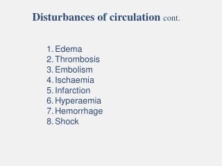
Disturbances of circulation cont. Edema Thrombosis Embolism Ischaemia Infarction Hyperaemia Hemorrhage Shock. 2. Thrombosis. The formation of solid mass of blood elements (blood coagulum) mainly fibrin and platelets inside the cardiovascular system during life.

E N D
Disturbances of circulation cont. Edema Thrombosis Embolism Ischaemia Infarction Hyperaemia Hemorrhage Shock
2. Thrombosis The formation of solid mass of blood elements (blood coagulum) mainly fibrin and platelets inside the cardiovascular system during life.
Mechanism of thrombosis 1-Deposition of platelets on the damaged endothelium of the blood vessels forming a white mass. Adherence of platelets lead to the release of ADP which cause further aggregation. 2- Aggregated platelets releases thromboplastin initiating coagulation by the following steps: Prothrombin (in plasma) +Thromboplastin(in platelets) Thrombin + Fibrinogen Fibrin forming mesh (white thrombus) at right angle to blood flow called zahn line. Mural thrombi 3- Fibrin mesh formed will entrap other platelets and RBCs forming the mixed thrombus. Ca++ Thrombin
Aggregation of platelets on damaged endothelium Release ADP Further aggregation Release thomboplastin Ca+2 Thrombin Prothrombin Forming mesh ┴ to blood flow which is called Zhan line Fibrinogen Fibrin The fibrin mesh formed will entrap other platelets and RBCs forming the mixed thrombus
Causes of Thrombosis 1- Roughness and stickiness of the endothelium of blood vessels. 2- Slowing of the blood flow (stasis) as in congestive heart failure, varicose and prolonged immobilization, this allows the platelets to come to the periphery. 3- Alteration of the blood constituents
Types of Thrombi 1- White Thrombus: Formed of platelets and fibrin as in case of vegetations on the heart valves of rheumatic fever patients. 2- Red Thrombus: Formed of RBCs and fibrin which occurs in the right side of the heart before death. A thrombus formed rapidly by the coagulation of stagnating blood. 3- Mixed Thrombus: Formed of all blood elements.
Sites of Thrombi 2- Heart: • Vegetations: formed over heart • valves following inflammation. • Mural: thrombi adherent to the • vessel walls • Agonal: A heart clot formed during the act of dying after prolonged heart failure 3- Veins: Two types are known • Thrombophlebitis(septic thrombus): occur secondary to inflammations caused by pyogenic bacteria. • Phlebothrombosis: occur due to slowing of the venous return as in varicose veins. 1- Arteries: Results from arteriosclerosis.
Sites of Thrombi Thrombophlebitis Phlebothrombosis
Fate of thrombi 1- Resolution 2- Organization 3- New blood vessels may be formed within the thrombus 4- Fragmentation.
3. Embolism It is an insoluble physical mass circulating through the blood until impacted in small blood vessel resulting in either i- Ischaemiaand infarctionof the organ supplied with the occluded artery. ii- Pyaemic abscessif the embolus is septic.
Types of emboli 1-Thromboemboli (veins, arteries, LS heart or aorta, RS heart) 2- Fat Emboli:
Types of emboli cont. • 3- Air or gas Emboli: • lung surgical operation,. • during delivery (rapture of uterine vein) • because of injecting air into uterus in criminal abortion, • faulty blood transfusion, • when pilots or divers are shifted from high to reduced pressure, the dissolved nitrogen forms bubbles • injury of neck vein.
Types of emboli cont. 4- Amniotic fluid: 5- Infective Emboli: 6- Tumor Emboli: 7- Foreign body, e.g. Bullet.
4. Ischaemia • It is the inadequate arterial blood supply to an organ due to obstruction of the blood flow with:- 1- Thrombus. 2- Embolus. 3- Thickening of arterial walls (arteriosclerosis). 4- Prolonged spasm of the artery muscle. 5- Pressure by a tumor on an artery. 6- Twisting or strangulation as in case of intestine. • If the site of occurrence has good collateral circulation, nothing will occur. • If it has bad collateral circulation the ischaemic tissue will die NECROSIS or degenerate if the occlusion is gradual.
Coronary artiers Middle cerebral arteries Small arteries of the limbs and digits 5. Infarction It is a localized area of ischaemicnecrosis, produced by the sudden and complete occlusion of either the arterial supply or the venous drainage. Infarction is more common in organs with bad collateral circulation such as brain, heart, spleen and kidney.
The necrotic area is opaque white and raised and irritates the surrounding living tissue leading to dark, red zone around it (dilated capillaries). The size of the infarct area depends on the size of the occluded artery and usually appear pyramidal in shape with its apex toward the occluded artery and the base to the surface of the infarct organ.
Cerebral infarction The characteristic cytological change, is ischaemic coagulative necrosis of the affected tissue, except the brain infarcts that is liquefactive.
6. Hyperaemia Increased blood due to vascular dilatation 1- Active Hyperaemia: Local increase of blood due to active dilatation of arterioles and capillaries. i- Physiologic ii- Pathologic
2- Passive Hyperaemia: (congestion) Increased blood due to passive dilatation of veins caused by venous obstruction. • Types of congestion a- Local venous congestion • Thrombus in vein (acute local congestion). • Pressure by tumor or bandage (acute local congestion). • Liver cirrhosis which causes intrahepatic obstruction of portal blood (chronic local congestion). b- General venous congestion • The obstruction is central (heart or lung). The general symptoms are cyanosis, dyspnea, pulmonary edema and generalized edema. The lungs and kidney become enlarged, heavier and dark brown in color.
7. Hemorrhage It is the escape or loss of blood from vessels due to their rupture. Causes of hemorrhage 1- Rupture of the vessel due to injury. 2- Hypertension. 3- Arteriosclerosis. 4- Peptic ulcer or esophagus varices. 5- Bacterial infection. 6-Vitamin K deficiency. 7- Hereditary in family e.g. hemophilia
Types of hemorrhage i 1- External Hemorrhage i- Epistaxis: ii- Hemoptysis: iii- Hematemesis: iv-Metorrhagia: uterine bleeding at irregular intervals, particularly between the expected menstrual periods. v- Menorrhagia: abnormally heavy and prolonged menstrual period at regular intervals vi- Meleana:obscure gastrointestinal haemorrhage iii ii
Types of hemorrhage cont. 2- Internal hemorrhage: i- Hemo thorax: bleeding in pleura. ii- Hemo pericardium: bleeding the pericardium. iii- Hemo peritoneum: bleeding in the peritoneum. iv-Interstitial Hemorrhage: Petechia, Echymosis, Hematoma ii i iii iv
Types of hemorrhage cont. iv- Interstitial Hemorrhage: Petechia, Echymosis, Hematoma • Petechia red or purple spot on the body, caused by a minor hemorrhage (broken capillary blood vesseles) • Echymosisthe escape of blood into the tissues from ruptured blood vessels • Hematoma a localized collection of blood outside the blood vessels, usually in liquid form within the tissue Petechia Echymosis Hematoma
Natural arrest of hemorrhage: (how body stop hemorrhage naturalluy) 1- Contraction and retraction of the blood vessel to narrow their lumen. 2- Formation of blood clot to seal the blood vessel. 3- Decrease in blood pressure and activation of vasomotor center. Signs of hemorrhage 1- Cold skin. 2- Thirst. 3- Hypoxia in vital organs. 4- Decreased blood pressure. 5- Faintness. 6- Severe hemorrhage results in shockand may be death.
8. Shock It is an acute persisting deficiency of blood flow in the peripheral vascular bed. Signs: Pale face, cold skin, rapid pulse, sweating, shallow respiration and low blood pressure 1- Primary shock It is neurogenic or psychic that immediately follows sever pain or emotion due to widespread vasodilatation and decrease in blood pressure leading to cerebral anemia and coma. It is usually reversible. 2- Secondary shock It is an acute circulatory failure and develops few hours after injury. It may be reversible or irreversible. It occurs after primary shock, severe acute inflammation, severe burn, continuous vomiting and diarrhea.