Stroke Rehabilitation
3.26k likes | 12.91k Views
Stroke Rehabilitation. Nursing implications. Learning objectives. At the end of this presentation the learner will: Understand the pattern of deficits for hemispheric, brain stem, and cerebellar CVAs. Understand the key nursing implications of care for a left and right hemiplegia.

Stroke Rehabilitation
E N D
Presentation Transcript
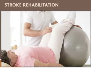
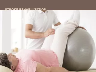
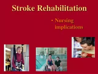
Stroke Rehabilitation • Nursing implications
Learning objectives • At the end of this presentation the learner will: • Understand the pattern of deficits for hemispheric, brain stem, and cerebellar CVAs. • Understand the key nursing implications of care for a left and right hemiplegia. • Understand the nursing care implications for common CVA deficits, aphasia, neglect, impaired sensory processing, motor, and visual field deficits.
Stroke: Definition Stroke is clinically defined as a neurologic syndrome characterized by acute disruption of blood flow to an area of the brain, and corresponding onset of neurologic deficits related to the concerned area of the brain Nurs Clin N Am 2002;37:35-57
Stroke: Classification Ischemic stroke: Account for 80%. Results from occlusion in a blood vessel supplying the brain • Thrombotic: Occlusion due to atherothrombosis of small/large vessels supplying the brain with blood • Embolic: Occlusion due to embolus arising either from heart (e.g. atrial fibrillation, valvular disease, PFO) or another blood vessel (DVT)
Classification Hemorrhagic stroke: Account for 20%. Results from rupture of blood vessels leading to bleeding in brain • Intracerebral: Bleeding within the brain due to rupture of small blood vessels. Occurs mainly due to high blood pressure • Subarachnoid: Bleeding around the brain; commonest cause is rupture of aneurysm.Other causes: Head injury secondary to trauma or fall
Hemispheric Expression of the stroke • Motor and sensory deficits are found on the side OPPOSITE to the affected side of the brain • Visual field deficits are also found on the side OPPOSITE to the affected side of the brain • Horizontal gaze is also affected in the direction OPPOSITE to the affected side of the brain • Because the eye can’t move to the opposite side, it actually appears to be looking AT the affected side of the brain in hemispheric strokes
Left(Dominant) Hemisphere Typical Signs: Right Side Weakness and Aphasia Right Visual Field Deficit Aphasia Left Gaze Preference (in hemispheric stroke, looks TOWARD the side of the injury) Right Hemiparesis Right Hemisensory Loss Hemiparesis: weakness or partial paralysis Hemiplegia: paralysis
Aphasia • In right hand dominant people, the speech center of the brain is found in the left hemisphere • So left hemispheric stroke is the most likely cause of aphasia in most people • HOWEVER, some left hand dominant people have their speech centers on the right side of the brain, so they may present with right hemispheric stroke symptoms and aphasia
Expressive aphasia (motor or Broca’s) • difficulty in selecting, organizing and initiating speech • speech is slow, hesitant and labored- short phrases or single words • Receptive aphasia (sensory or Wernicke’s) • impaired auditory comprehension and feedback, unable to monitor and correct speech • Speech may be of normal rate and grammar intact, however unaware of and unable to correct mistakes; may substitute a group of sounds, words or syllables • Global aphasia • nonfluent speech with poor comprehension and limited ability to name objects or repeat words
Right(Nondominant) Hemisphere Typical Signs: Left Side Weakness Left Hemi-inattention (Neglect) Left Visual Field Deficit Right Gaze Preference (in hemispheric stroke, looks TOWARD the side of the injury) Left Hemiparesis Left Hemisensory Loss
Hemi-inattention or “Neglect” • Patients with neglect tend not to acknowledge anything about the affected side of their body • “People who experience damage to the right parietal lobe sometimes show a fascinating condition called hemi-inattention. When this occurs, the person is unable to attend to the left side of the body and the world. A person with hemi-inattention may shave or apply makeup only to the right side of the face. While dressing, he or she may put a shirt on the right arm but leave the left side of the shirt hanging behind the body. The person may eat from only the right side of the plate, not noticing the food on the left side. This condition is not due to visual problems or the loss of sensation on the left side of the body, but is a deficit in the ability to direct attention to the left side of the body and the world.” (Psychobiology, Salem Press)
Hemi-inattention or “Neglect” • The most common form of neglect is neglect of the left side of the body due to a right hemispheric lesion • If a patient appears not to acknowledge your presence from one side of the body, try changing sides to rule out hemi-neglect • Patients can often eventually totally recover from hemi-inattention deficits
Do you think you will have difficulty? “None”Task is performed Did you have any difficulty? “None”
Failure to recognize side of body contralateral to injury • May not bathe contralateral side of body or shave contralateral side of face • Deny own limbs • Objects in contralateral visual field ignored
Right sided paralysis Communication deficits Aphasia- expressive, receptive & Global aphasia Loss of problem solving skills Right visual field deficit Emotional Lability Decreased organizational skills and initiation Disoriented to time & place Perseverative movements & phrases Left CVA
Vision-Unable to discriminate words & letters or read. Deficits in right visual field Behavior-slow, cautious, anxious when attempting new task Depression or catastrophic response to illness, sense of guilt, Emotional Lability Feeling of worthlessness, worries over future, is quick to anger & becomes frustrated easily. Left CVA
Right CVA • Left sided Paralysis • Left visual field deficits • Agnosia – inability to recognize familiar objects (keys, pen, persons) • Poor Judgement • Impulsive behavior • Denial of deficit • Easily distracted • Unilateral neglect
Right CVA • Visual spatial deficits • Neglect in left visual field, loss of depth perception • Impulsive behavior – unaware of deficits • Confabulates –Euphoric • Constant Smile • Poor judgement • Over estimates abilities
Brainstem Typical Signs: Bilateral Abnormalities Crossed Signs (1 side of face and contralateral body) Quadriparesis Sensory Loss in All 4 Limbs Hemiparesis Hemisensory Loss
Cranial nerve signs suggest localization to (and within) the brainstem
Brainstem Typical Signs: Cranial Nerve and Other Deficits Vertigo, Tinnitus Dizziness Decreased LOC Nausea, Vomiting Hiccups, Abnormal Respirations Eye Movement Abnormalities: Diplopia Dysconjugate Gaze Gaze Palsy (horizontal gaze deficit or gaze preference) Nystagmus Oropharyngeal Weakness: Dysarthria (speaking), Dysphagia (swallowing)
Cerebellum Typical Signs: Lack of Coordination Ipsilateral (same side) Limb Ataxia (dyscoordination) Truncal or Gait Ataxia (imbalance) Tremors, or Limb Ataxia, result from lack of coordination of opposing muscle groups (flexors vs. extensors), causing the muscle groups to fight each other
REHABILITATION Restoration of a disabled person to maximum independence by developing his/her residual capacities.
”Spontaneous” recovery • ”Spontaneous” recovery from, e.g., stroke • Quick recovery of functions during the first three months after injury • Slower recovery thereafter, but can improve over years if they keep working on it
Theories of Recovery • Resolution of harmful factors • Reduced edema, resorption of toxins, increased circulation • Neuroplasticity • Collateral sprouting - From intact cells to denervated region after some or all input has been destroyed • Unmasking of neural pathways and synapses not normally used • Can be altered by drugs, environmental conditions, electrical stimulation
Figure 5.25 Collateral sprouting A surviving axon grows a new branch to replace the synapses left vacant by a damaged axon.
Adult Plasticity and Regeneration The brain has an amazing ability to reorganize itself rapidly through new pathways and connections . • Through Practice: • Motor regions • After damage or injury • Undamaged neurons make new connections and take over functionality or establish new functions • But requires stimulation • Stimulation is a standard technique for stroke survivor in rehabilitation
Cardinal Principles of Rehab • E: Early Treatment • A: Activity Strengthens • S: Stress Abilities, NOT disabilities • T: Treat total patient • Treat adults as adults!
Essential nursing competencies Protect, maintain, restore and promote the health of individuals and the command of their vital physical and mental functions taking into account the personality of each person and his psychological, social, economic and cultural characteristics.
Unilateral Neglect • This syndrome is most commonly seen with right cerebral stroke. • Teach client to: • Observe safety measures. • Touch and use both sides of the body. • Use scanning technique of turning the head from side to side to expand the visual field
Nursing Intervention for Stroke Deficits Motor Positioning, alignment, ROM Provide alternative communication Test reflexes before offering nourishment; elevate head Speech consultation Hemiparesis or hemiplegia Dysarthria Dysphagia
Sensory Deficits Teach patient to check body parts visually Protect involved area; accept pt.'s perception; position pt. to face involved area Controlamt. of change in schedule; reorient Correctmisuse of object; demonstrate correct use Correct misinformation Place equipment where pt. can see it Reduce distraction Phraserequests without R/L designation
Language and Cognitive Deficits Memory loss Short attention span Distractibility Poor judgment Inability to transfer learning Inability to calculate, reason, or think abstractly Provide information Divide activities in small segments Control distractions Protect pt. from injury Repeat, Repeat, Repeat Keep expectations realistic & keep it simple Expressive Aphasia Speak clearly, use tactile cues & gestures. Receptive Aphasia Patience!!!! Global Aphasia Mime techniques
Impaired Mobility and Self-Care • Interventions include: ROM exercises for the involved extremities Change of client’s position frequently Prevention of deep vein thrombosis Therapy focused on ADLs Reinforce specific techniques learned in therapy
Urinary & Bowel Incontinence • Altered level of consciousness may cause incontinence or impaired innervation, or an inability to communicate. • Develop a bladder and bowel training program.
Bladder Retraining • Diagnosis • Rule out reversible causes-UTI’s, BPH , Meds • Post-void residuals-Retention • Urodynamic studies • Treatment • Timed toileting – use toilet or commode to promote optimal emptying of bladder, men should stand to void if able • Fluid restriction after dinner • External catheters • Intermittent or indwelling catheterization • Medications
Bowel Retraining • Bowel Dysfunction • Causes • Disinhibition of reflex emptying mechanisms, sensation or cognitive impairments • Prevention & Treatment • Diet: adequate fluids, fiber • Toileting after meals (gastrocolic reflex) • Medications: stool softeners, bowel stimulants, suppositories, enemas • Use toilet or commode chair for best results if possible • Persistent bowel incontinence >4 weeks usually poor functional predictor
Medical Complications • Pressure Sores • Preventive Strategies • Nutrition • Hydration • Incontinence care • Specialty Mattresses • Heel protector boots • Positioning and turning • Pressure relief
Medical Complications • Deep Venous Thrombosis (DVT) • Incidence • Up to 20% to 75% of stroke survivors • Preventive • Stockings • Thigh-high TED’s • Pneumatic compression/SCD’s • Subcutaneous heparin or Lovenox, • Treatment • Heparin, Lovenox • Warfarin
Medical Complications • Shoulder Pain • Causes • Impaired passive range of motion • Adhesive capsulitis • Neuropathy • Chronic regional pain syndrome (CRPS), RSD (Reflexive Sympathetic Dystrophy) or Shoulder Hand Syndrome • Shoulder trauma • Bursitis Tendinitis • Rotator cuff tear • Heterotropic ossification
Medical Complications • CRPS Type I Treatment for shoulder pain • Aggressive range of motion (ROM) • Pharmacologic agents • Nonsteroidal agents • Antidepressants • Local injections • Corticosteroids • Gabapentin • Sympathetic blocks • eTENS
Medical Complications • Shoulder Subluxation • Pathogenesis not well understood • Supraspinatus weakness implicated • Treatments • Shoulder supports • Functional electrical stimulation (FES) • Arm boards • Overhead slings • Never lift under hemiparetic arm during transfers or bed mobility
Medical Complications • Spasticity • Treatment • Goals • Prevention of deformities • Tone inhibition • Modalities • Orthoses • Static activities • Inhibitory • Dynamic activities • Surgery • Muscle release • Tendon lengthening
Medical Complications • Spasticity Treatment Medications • Systemic • Dantrolene • Clonidine • Tizanidine • Oral Baclofen • Neurolytic Agents • Phenol or denatured alcohol blocks • Botulinum toxin • Intrathecal • Baclofen pump
Medical Complications • Dysphagia • Occurrence • Up to one·third of stroke survivors • Complications • Malnutrition /Dehydration • Aspiration Pneumonia • Aspiration Symptoms • Dysphonia, wet voice quality • Decreased gag reflex • Decreased cough reflex • Elevated temp, abnormal lung sounds
Dysphagia • Interventions include: • Assessment of client’s ability to swallow via Speech Therapy evaluation, video fluoroscopy, fiberoptic laryngoscopy • Client head positioning to facilitate the process of swallowing before feeding • Appropriate diet for the client, including modified textures of foods and fluids • Utilization of compensatory strategies during feeding (double swallow, chin tuck, use of straws etc.)