

E N D
Presentation Transcript
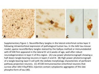
Supplementary Figure 1. Neurofibrillary tangles in the lateral entorhinal cortex layer II following intraentorhinal expression of pathological human tau. In the AAV-tau mouse model, sparse neurofibrillary tangles stained by the Gallyas method or immunolabeled with AT100 first appeared in the lateral EC at 6 weeks of age, well after robust neurodegeneration in layer II of this region. (A) Low power photomicrograph showing a half dozen tangle-bearing neurons in layers II and III. (B) High power photomicrograph of a tangle-bearing layer II cell with the stellate morphology characteristic of perforant pathway projection neurons. (C) AT100 immunoreactive entorhinal neurons that survive after AAV-hTauP301L injection contain cytoplasmic aggregates of this late phospho-form of tau (40).