Download
1 / 21
300 likes | 748 Views
CT and MR Imaging of Abdominal Aortic Aneurysm. Rocio Cabrera Guillaume Lemaitre Mojdeh Rastgoo. Presentation Outline. Introduction to Abdominal Aortic Aneruysms Computed Tomography of AAA Imaging Technique Image Processing Level Set Methods Active Shape Models

E N D
CT and MR Imaging of Abdominal AorticAneurysm Rocio Cabrera Guillaume Lemaitre MojdehRastgoo
PresentationOutline • Introductionto Abdominal AorticAneruysms • ComputedTomography of AAA • ImagingTechnique • ImageProcessing • Level Set Methods • Active ShapeModels • MagneticResonance of AAA • ImagingTechniques • Galodinium-enhancement • Diffusionweighted • ImageProcessing • MarkovianMethod • Graph-TheoreticApproach • Conclusions Biological Basis of Medical Imaging - CT and MR Imaging for Abdominal Aortic Aneurysms
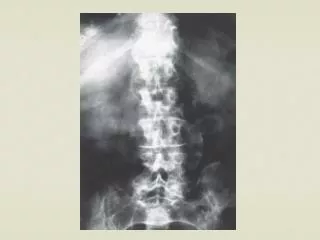
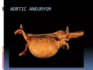
IntroductionWhatisan Abdominal AorticAneurysm? • Aneurysm • Vascular pathology consisting of an irreversible dilation of a segment of a blood vessel • Abdominal aorta • Continuation of the thoracic aorta and begins at the level of the diaphragm • Largest artery in the abdominal cavity • Abdominal Aortic Aneurysm • Acceptedcriterion: 50% increasein vesseldiameter Biological Basis of Medical Imaging - CT and MR Imaging for Abdominal Aortic Aneurysms
CT Imaging CT has been used widely in AAA CT Imaging speed CT provides detailed quality for better analysis of aneurysm and adjacent arteries Detailed information of aorta and its branches for 3D reconstruction Flexible to different post processing methods Appreciated in surgical planning Biological Basis of Medical Imaging - CT and MR Imaging for Abdominal Aortic Aneurysms
CTImageProcessingAAA Segmentationthroughlevel set method • Level Set Method (LSM) is a numerical method for tracking interface a shapes . More specifically for shape varying objects plane Level set function Biological Basis of Medical Imaging - CT and MR Imaging for Abdominal Aortic Aneurysms
CTImageProcessingAAA Segmentationthroughlevel set method 1 - Segmentation the volumes Defining the mesh on the object (Level Set function ) Force at mesh point (x,y) Image force based on the Gaussian derivative filter Curvature force (x’ ,y’) Advection term = 1 Updating the mesh values using the Speed function Multi resolution Analysis – LR( half) volume Top – Down , Narrow band update restricted to the zero level set Scaling up the result Repeating the algorithm on full data set 2 - 3D reconstruction – Using Marching cubes Biological Basis of Medical Imaging - CT and MR Imaging for Abdominal Aortic Aneurysms
CTImageProcessingAAA Segmentationthroughlevel set method • Advantage: • Level set method has the advantage to provide more accurate results specially in segmenting the small details • Disadvantage • It has a very high computational cost • Suggestions • Combination of the methods , while level set can be used to improve the initial segmentation results Biological Basis of Medical Imaging - CT and MR Imaging for Abdominal Aortic Aneurysms
CTImageProcessingAAA Segmentationusing Active ShapeModel • Active shape model (Smart Snake) was developed by Cootes et al. in order to over come the problems with snake segmentation • Active counter model (Snake) segmentation depends on the initial snake • Active counter model is not capable to deal with the occluded objects • In Medical Imaging ASM is applied on the combination of shapes and gray level sets Biological Basis of Medical Imaging - CT and MR Imaging for Abdominal Aortic Aneurysms
CTImageProcessingAAA Segmentationusing Active ShapeModel Shape Modeling Shape alignment Statistical Computations PCA Aligning the images in the same reference axes , using Procrustes Analysis Procrustes Analysis minimize the distance between reference shape and each shape in the dataset Modeling the shape variations Computation of the mean shape Computation of the scatter matrix Sorting the eigenvectors and keeping the first k eigenvectors , based on the largest eigenvalues Eigen decomposition of the shapes where , Value of k is based on Biological Basis of Medical Imaging - CT and MR Imaging for Abdominal Aortic Aneurysms
CTImageProcessingAAA Segmentationusing Active ShapeModel Shape Modeling M. Bruijne ,B. van Ginneken, M. A. Viergever, W. J. Nieesen, « Interactive Segmentation of Abdominal Aortic Aneurysms in CTA Images », 2004 Biological Basis of Medical Imaging - CT and MR Imaging for Abdominal Aortic Aneurysms
CTImageProcessingAAA Segmentationusing Active ShapeModel Grey level Appearance Modeling M. Bruijne ,B. van Ginneken, M. A. Viergever, W. J. Nieesen, « Interactive Segmentation of Abdominal Aortic Aneurysms in CTA Images », 2004 Sum of absolute difference between the reference and sample image over several resolutions The sum is performed for each landmarks for a defined window size Biological Basis of Medical Imaging - CT and MR Imaging for Abdominal Aortic Aneurysms
CTImageProcessingAAA Segmentationusing Active ShapeModel Model Fitting M. Bruijne ,B. van Ginneken, M. A. Viergever, W. J. Nieesen, « Interactive Segmentation of Abdominal Aortic Aneurysms in CTA Images », 2004 First slice manually Initialized while for the others previous counter was considered as initialization Performing the multiresolution analysis for higher accuracy Biological Basis of Medical Imaging - CT and MR Imaging for Abdominal Aortic Aneurysms
MR Imaging of AAAAlternate MR Imaging Approaches Gadolinium-enhanced MRI Diffusion Weighted MRI • Gadolinium injection (paramagnetic CA) • Shortens the T1 relaxation time of blood, distinguishing it from its surroundings • No known side effects nor nephrotoxicity • Prince et al. [2] reported an agreement in measured AAA size in CT, MR and US • Orta et al. [1] reported its use to diagnose inflammatory AAA • Hyper-intensity surrounding the aorta • Region ADC = 1.24 x 10⁻² mm2/s • ADC consistent with a restricted diffusion due to inflammation Biological Basis of Medical Imaging - CT and MR Imaging for Abdominal Aortic Aneurysms
MR ImageProcessingMarkovian-Active ContourSegmentation • X – 2D [MxN] random field that models the segmentation labels • Y – 2D [MxN] random field that models the input image • s – site (pixel) located at position (i,j) • MAP – Maximum A Posteriori Probability, through the Bayes Rule • Assuming Gibbsian distributions A pixel s will switch classes if and only if at least one of its neighbors has already been assigned the new class label Likelihood Energy. Natural Logarithm of a Gaussian Biological Basis of Medical Imaging - CT and MR Imaging for Abdominal Aortic Aneurysms
MR Image ProcessingMarkovian-Active Contour Segmentation • Direct extension to 3D and 4D • 3D – Neighbouring sites in k+1 and k-1 images • 4D – Neighbouring sites in t+1 and t-1 time frames • AAA reconstruction from MRI • Initialization done by expert hand • Seed growth until convergence • 4D segmentation results Biological Basis of Medical Imaging - CT and MR Imaging for Abdominal Aortic Aneurysms
MR Image ProcessingGraph-TheoreticSegmentation • Aortic Surface Pre-segmentation • Fast marching level set method used to compute {appSt}tє[0,N-1] • Centerline Extraction • Centerline determined from each approximate surface by skeletonization • Accurate Surface Segmentation • Weighted graph G = (V; E) • V – node set of image pixels • E – arc set of neighbourhood system • Every arc ‹vi, vj› є E has a cost • Graph-cuts aim to partition a weighted graph into 2 disjoint subsets • Minimize the cost function ε(f) • Appropriate design of a energy function, a minimum s-t cut can segment a region of interest in an image. Biological Basis of Medical Imaging - CT and MR Imaging for Abdominal Aortic Aneurysms
Conclusions • Strong interest in exploiting the capabilities of the medical imaging modalities to diagnose AAA • Choice of the imaging modality • CT • Modality of choice in most institutions • Appropriate for emergency patients • Use of an iodinated contrast medium and ionizing radiation • MR • Appropriate to detect inflammatory AAA • Does not employ ionizing radiation • Contrast agent is appropriate for patients with renal insufficiency • Image processing techniques • 2 CT and 2 MR methods have been presented • Most methods rely on segmentation of the aorta and measurement of the vessel diameter • Each methods could be extended to the other modality, but not much research has been done on it • It would be interesting to perform a study in which the several segmentation methods are used on CT and MR in order to evaluate the methods and the imaging modalities in a better way. Biological Basis of Medical Imaging - CT and MR Imaging for Abdominal Aortic Aneurysms
References [1] Orta, K. and Kilickesmez, O. Clear Depiction of Inflammatory Abdominal Aortic Aneurysm with Diffusion-Weighted Magnetic Resonance Imaging. Cardiovascular and Interventional Radiology. (2010) 33:379-382. [2] Prince, M. et al. Gadolinium-enhanced Magnetic Resonance Angiography of Abdominal Aortic Aneurysms. Journal of Vascular Surgery. (1995) Volume 21. Number 4. [3] Jodoin, P. et al. Markovian Method for 2D, 3D and 4D segmentation of MRI. (2008) 15th IEEE International Conference on Image Processing. [4] Sonka, M. et al. Early Detection of Aortic Aneurysm Risk from 4D MR Image Data. (2006) Computers in Cardiology. [5] Kang Li et al. Optimal Surface Segmentation in Volumetric Images - A Graph-Theoretic Approach (2006) IEEE Transactions on Pattern Analysis and Machine Intelligence. Volume 28. Number 1. [6] Crawford, C. et al. Abdominal Aortic Aneurysm: An Illustrated Narrative Review. (2002) Journal of Manipulative and Physiological Therapeutics. Volume 26. Number 3. [7] Marleen de Bruijne and Bram van Ginneken and Max A. Viergever and Wiro J. Niessen. Interactive Segmentation of Abdominal Aortic Aneurysms in CTA Images. 2004. [8] TF. Cootes and A. Hill and C.J.Taylor and J.Haslam and Manchester M Pt. The Use of Active Shape Models For Locating Structures in Medical Images. 1994. [9]T.F. Cootes and C.J. Taylor and Manchester M Pt. Statistical Models of Appearance for Computer Vision. 2000. [10] Cootes, T. F. and Taylor, C. J. and Cooper, D. H. and Graham, J. Active shape models - their training and application. Comput. Vis. Image Underst. 1995. Volume 61. Issue 1. Pages 38-59. [11] A. Hill and A. Thornham and C. J. Taylor. Model-Based Interpretation of 3D Medical Images. In British Machine Vision Conference. 1993. BMVA Press. [12] Michael Kass and Andrew Witkin and DemetriTerzopoulos. Snakes: Active contour models. International Journal of Computer Vision. 1988. Volume 1. Number 4. [13] Derek Magee and Andrew Bulpitt and Elizabeth Berry. Level Set Methods for the 3D Segmentation of CT Images of Abdominal. [14] Steven C. Mitchell and Boudewijn P. F. Lelieveldt and Rob J. van derGeest and Hans G. Bosch and Johan H. C. Reiber and Milan and Milan Sonka. Multistage Hybrid Active Appearance Model Matching: Segmentation of Left and Right Ventricles in Cardiac MR Images. IEEE Transactions on Medical Imaging. 2001. Volume 20. [15] Wikipedia. Level Set Method. December 2010. $http://en.wikipedia.org/wiki/Level\_set\_method$ [16] Wikipedia. Isosurface. December 2010. $http://en.wikipedia.org/wiki/Isosurface$ [17] Wikipedia. Marching Cubes. December 2010. $http://en.wikipedia.org/wiki/Marching\_cubes$ [18] George G. Hartnell. FRCR, FACC. Imaging of Aortic Aneurysm and Dissection: CT and MRI. Journal of Theoretical Imaging. 2001. Biological Basis of Medical Imaging - CT and MR Imaging for Abdominal Aortic Aneurysms
Appendix - AAA Segmentation through level set method • Defining static and evenly spaced mesh in the image using • Mesh values are updated using the speed function • Speed function F depends on : • Advection term (constant value) • Curve term based on zero level set • Image term (Based on the edges) Updating the curve and Image terms based on nearest neighbor in the zero level set !! Computationally expensive Reducing the computational cost by updating the mesh restricted to the zero level set area ; Narrow band with the level set method Biological Basis of Medical Imaging - CT and MR Imaging for Abdominal Aortic Aneurysms
IntroductionPathogenesis of an Abdominal AorticAneurysm Environment Atherosclerosis Smoking Inflamation GeneticFactors Elastaseactivityincrease vs. Inhibitiondecrease ElastinDestruction Failure of Elastin Collagenremodelling Hypertension Increased load oncollagen Ageing Failure of Collagen Alteration in vesselgeometry AneurysmalDilation Rupture • Elastin • Responsible for the elastic recoil of the arteries due to the pulsatile blood flow • Degradation of elastinfibres will shift load to collagen fibres • Elasticity decrease • Diameter increase • Aortic rupture • Genetic and environmental factors contribute to AAA development • Great clinicalimportanceto determine aorticdiameter Biological Basis of Medical Imaging - CT and MR Imaging for Abdominal Aortic Aneurysms
MR Imaging of AAATraditional MRI limitations on Vascular Imaging • Traditional MRI • Widely used to retrieve anatomical and physiological information of patients • Vascular imaging limitations • Flow artifacts generated in different pathologies • Aneurysms – slow, swirling flow • Stenotic vessels – turbulent flow Biological Basis of Medical Imaging - CT and MR Imaging for Abdominal Aortic Aneurysms