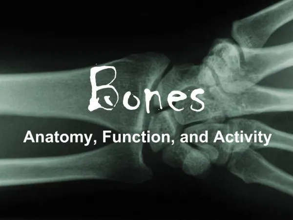
Dem Bones
Dem Bones. Factoids. The human hand has 27 bones; your face has 14! The longest bone in your body? Your thigh bone, the femur -- it' s about 1/4 of your height. The smallest is the stirrup bone in the ear which can measure 1/10 of an inch.

Dem Bones
E N D
Presentation Transcript
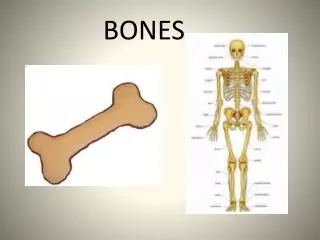
Factoids • The human hand has 27 bones; your face has 14! • The longest bone in your body? Your thigh bone, the femur -- it' s about 1/4 of your height. The smallest is the stirrup bone in the ear which can measure 1/10 of an inch. • Did you know that humans and giraffes have the same number of bones in their necks? Giraffe neck vertebrae are just much, much longer! • You have over 230 moveable and semi-moveable joints in your body.
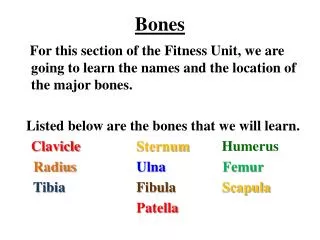
Skeletal System • Bones are made of several tissues • Primarily made of collagen and hydroxyapatite - Ca10(PO4)6(OH)2 • 206 bones in the human body (over 300 at birth).
Functions of Skeletal System • SUPPORT: Hard framework that supports and anchors the soft organs of the body. • PROTECTION: Surrounds organs such as the brain and spinal cord. • MOVEMENT: Allows for muscle attachment therefore the bones are used as levers. • STORAGE: Minerals and lipids are stored within bone material. • BLOOD CELL FORMATION: The bone marrow is responsible for blood cell production.
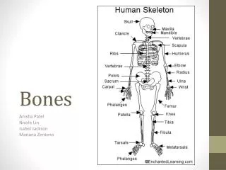
Skeletal System Organization • Axial skeleton • Skull and bones that support it • Includes vertebra and ribs • 80 bones • head • neck • trunk
Skeletal System Organization • Appendicular skeleton • Limbs • 126 bones • upper limbs • lower limbs • pectoral girdle • pelvic girdle
Vertebral column First 7 - cervical Next 12 - thoracic Last 5 – lumbar Maxilla Mandible Illium Ischium Pubis Ribs (12 pairs) First 7 - true Next 5 - false (Last 2 - floating) Calcaneus Talus
Bone Classification • Flat Bones (c) • cranium, ilium, sternum, rib cage, sacrum and scapula • Sesamoid (Round) Bones (e) - patella • Irregular Bones (d) • vertebrae, sacrum, hyoid Short Bones (b) - bones of the wrist and ankle Long Bones (a) • femur, tibia, fibula, humerus, radius, and ulna , metacarpals, metatarsals, and phalanges
Features of a Long Bone: Epiphysis: Ends of the bone. Diaphysis: The shaft of the bone which surrounds the medullary cavity. Articular Cartilage: Cushions the ends of the bones and allows for smooth movement. Epiphyseal Plate: Areas made of cartilage allowing for the growth of the bone.
What are the following depressions & openings? Foramen Fossa Meatus
What are the following processes? Tuberosity Head Facet Condyle
Bone Structure • Periosteum – hard outer covering • Cells for growth and repair • Compact bone – hard strong layer • Bone cells, blood vessels, protein with Ca and P • Spongy bone – at ends of long bones • Has small open spaces to lighten weight • Marrow cavity – hollow in middle of long bones
Bone Marrow • Red marrow – produces blood cells and clotting factors • Found in humerus, femur, sternum, ribs, vertebrae, pelvis • Produces RBC 2 million per second • Yellow marrow – stores fat • Found in many bones
Hematopoiesis • Occurs in cavities with red marrow • Medullary Cavity • In children contains read marrow and is hematopoietic • In adults the red marrow is replaced by fat – yellow marrow (not hematopoietic) • Hematopoiesis in Adults • Red marrow in spaces of spongy bone • Head of femur and humerus • Some flat bones: sternum, pelvic bone
Bones are composed of connective tissue, chemicals, and fats Solid outer layer - compact bone Composed of osteons An inner layer of spongy bone a honeycomb of flat, needle-like projections called trabeculae. Bone Structure Above: Note the relationship btwn the compact and spongy bone. Below:Close up of spongy bone.
Volkmann’s canals Perpendicular to the haversian canals. Connect the blood and nerve supply in the periosteumto those in the haversian canalsand the medullary cavity. Compact Bone • Haversian canals • allow the passage of blood vessels, lymphatic vessels, and nerve fibers. • Surrounded by layers of bone called a lamella. osteon
Bone Cells • Osteoblasts • Bone building cells • Synthesize and secrete collogen fibers and other organic components of the bone matrix • Initiate calcification • Found in the periosteum and the endosteum • Ossification • Formation of bone by osteoblasts. • Cells surround themselves by matrix. osteoblasts Bone matrix
Osteocytes. Mature bone cells. Osteoblasts that have become trapped by the secretion of matrix. Responsible for maintaining the bone tissue Lacunae spaces occupied by osteocyte cell body Canaliculi canals that allow for nutrient filled liquid to fill the lacunae Bone Cells 6-22
Bone Cells • Osteoclasts • Cells that ecretes digestive enzymes to digest bone matrix • bone resorption • Concentrated in the endosteum. • On the side of the cell that faces the bone surface, • ruffled border. • Pumps out hydrogen ions • Create an acid environment that eats away at the matrix.
Bone Cells Why is there a depression underneath the osteoclast? What advantage might a ruffled border confer? What is the name of the third cell type shown here? What do you think the tan material represents? www.academic.pgcc.edu/~aimholtz/AandP/LectureNotes/ANP1_Lec/Skeletal/BoneTissue.ppt
Bone Development • Initial skeleton of cartilage in infants • Replaced with bone by osteoblasts • More than 300 bones at birth – fuse to 206 • Always growing and breaking down • Osteoblasts – form new bone cells • Osteoclasts – break bone cells down • Osteocytes – mature bone cells
Types of Fractures • green stick • fissured • comminuted • transverse • oblique • spiral
Fracture Repair • Hematoma- blood clot in space between edges of break • Fibrocartilage callus- begins tissue repair • Bony callus- osteoblasts produce trabeculae (structural support) of spongy bone and replace fibrocartilage • Remodeling- osteoblasts build new compact bone, osteoclasts build new medullary cavity
Broken Bones Regrowth of bone: • Spongy bone forms in first few days • Blood vessels regrow and spongy bone hardens • Full healing takes 1-2 months
Mechanics of Movement Tissues and Structures Involved Muscle Nerve Bone Cartilage What are Tendons &Ligaments? Typesof Joints Mechanicsof Joints
HOW DOES MOVEMENT HAPPEN? MusclesPULL on Tendons to Move Bones at Connections called JointsorArticulations
Tendons Tendons are structures that connect bone to muscle, muscle to muscle, or bone to bone • Made up of tendon tissue (connective tissue) • Can have various shapes • Typical is cord-like tendon of biceps Frolich, Human Anatomy, Mechanics of Movement
Ligaments • Ligaments connect bone-to-boneorreinforce joints--they are made up of tendonoustissueas well • Typical are knee ligaments Frolich, Human Anatomy, Mechanics of Movement
Usually, but not always allow for movement Formed from various connective tissues Fibrous Cartilaginous Synovial(most complex--typical limb joints) Functionsof joints Hold bones together Allow for mobility Ways joints are classified Functionally Structurally Joints Frolich, Human Anatomy, Mechanics of Movement
Functional Classification The amount of movement the joint allows • Synarthroses • immovable joints • Amphiarthroses • slightly moveable joints • Diarthroses • freely moveable joints Frolich, Human Anatomy, Mechanics of Movement
Structural Classification • Fibrous joints • Generally immovable • Fibrous tissue separate the boney region at the joint • Cartilaginous joints • Immovable or slightly moveable • Cartilage separates the boney regions at the joint • Synovial joints • Freely moveable • The boney regions of the joint are separated by a space Frolich, Human Anatomy, Mechanics of Movement
Fibrous Joints • Bones united by fibrous tissue – synarthroses or largely immovable. • Skull • Bones tightly bound by minimal fiber • Syndesmosis • Longer connecting fibers • Joint has more give Frolich, Human Anatomy, Mechanics of Movement
Cartilaginous Joints • Mostlyamphiarthrosis • Bones connected by cartilage • Slightly movable • Pubic symphysis • Intervertebral joints • Hyaline cartilage unites bones • Epiphyseal growth plates • Costal cartilage-sternum Frolich, Human Anatomy, Mechanics of Movement
Synovial Joints • Diarthroses – movable joint • Most common joint in the body • Articulating ends of bones are covered with hyaline cartilage • Enclosed by a capsule of fibrous connective tissue lined with synovial membranes • Joint cavity is filled withsynovialfluid for lubrication • Ligamentsreinforce the joint Frolich, Human Anatomy, Mechanics of Movement
Typical Synovial Joint Frolich, Human Anatomy, Mechanics of Movement
Structures Associated with the Synovial Joints • Bursae – flattened fibrous sacs • Lined withsynovial membranes • Filled withsynovial fluid • Not actually part of the joint • Tendon sheath • Elongated bursathat wraps around a tendon
Types of Synovial Joints The type of joint, in part, determines the range and direction of movement
Types of Synovial Joints The type of joint, in part, determines the range and direction of movement
Upper arm bone - humerus Capsule (ligaments) Synovial membrane Cartilage Cartilage Synovial fluid tendon Triceps muscle HOW DOES MOVEMENT HAPPEN? The elbow joint - a hinge joint allowing movement in 1 plane The Capsule. Holds the bones of the joint in place. The synovial membrane. Secretes synovial fluid The synovial fluid. Lubricates the movement of the cartilage surfaces against each other – reducing friction and preventing arthritis (inflammation and joint damage). Cartilage. Lubricates the movement of the cartilage surfaces against each other – reducing friction and preventing arthritis (inflammation and joint damage).
HOW DOES MOVEMENT HAPPEN? The elbow joint - how the bicep and triceps control movement When the biceps contracts the elbow joint flexes (its joint angle decreases). humerus biceps (flexor muscle), contracts When the triceps contracts the elbow joint extends (its joint angle increases). Triceps (extensor) relaxes The biceps and triceps are called antagonistic muscles because they have the opposite effect on the same joint. radius ulna Remember that for this to work properly the biceps must relax when the triceps contracts, and vice versa. THE BICEPS AND TRICEPS ARE AN ANTAGONISTIC PAIR