Download
1 / 83
840 likes | 1.05k Views
The Retina. Introduction - Images from the retina are analyzed for form/movement/color. For each point in the outside world, there is a corresponding image point on the retina in each of the two eyes. .

E N D
Introduction • - Images from the retina are analyzed for form/movement/color. • For each point in the outside world, there is a corresponding image point on the retina in each of the two eyes.
These images from the two eyes are then brought together and compared at higher stations in the visual pathway for extraction of information about depth. • - Development of retina: The prosencephalon protrudes laterally and enlarges to form primary optic vesicles.
These later invaginate to give double-walled optic cups, with a largely obliterated ventricular space between the two walls. • The outer wall becomes the pigment epithelium, while the inner wall becomes the neural retina. • The two are separated by the subretinal space, the residue of the ventricular cavity.
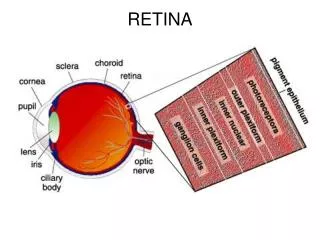
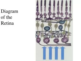
I. Structure of retina • - The retina is composed five main types of neurons, segregated in to three main layers: • 1) Outer nuclear layer: photoreceptors, no direct blood supply. • Instead, they receive nourishment from the choroidal circulation filtered thru the pigment epithelium.
2) Inner nuclear layer: bipolar (second order visual neurons), horizontal (lateral association neurons), amacrine (ditto), Mueller glial cell bodies • 3) Ganglion cell layer: retinal ganglion cells (third order visual neurons, who axons constitute the optic nerve). • - The outer and inner plexiform layers are sites of synapses between layers
II. Photoreceptors • - There are two types of photoreceptors • 1) rods: most sensitive in dim light, only one type • 2) cones: less sensitive to light, three types mediate color vision (peaks at blue, green and yellow).
- Distribution of photoreceptors is not uniform: fovea has high density of cones and few rods, periphery has more rods. • Blue sensitive cones are also absent from the center of the fovea. • - The fovea has the highest visual acuity, or spatial resolution of the visual space.
This is achieved by having thinner cones and more of them per unit space than elsewhere in the retina; with the layers of cells anterior to the receptors being pushed aside. • This clearing minimizes light scattering which tends to degrade the quality of the image.
- The structure of the photoreceptors: • o Outer segment: where light is absorbed and a neural signal is generated. • It contains an orderly stack of membranes in which is embedded the light sensitive visual pigment (rhodopsin in rods, and one of three cone pigments in cones). • In cones these membranes are continuous with the plasma membrane to form a highly convoluted surface membrane.
In cones these membranes are continuous with the plasma membrane to form a highly convoluted surface membrane. • In rods, the membranes are completely internalized to form a stack of flattened membranous discs.
The high density of pigment provides a high probability of absorption of an incident photon. • o Inner segment: contains the metabolic machinery of the cell, while the synaptic terminal forms chemical synaptic connections with bipolar and horizontal cells. • o Glu is the NT released by both rods and cones.
- Pigment epithelial cells serve to • (1) absorb light not absorbed by the rods/cones using melanin, • (2) phagocytoseshedded fragments of rods/cones, • (3) regenerate pigment by supplying frestchromophore to the bleached pigment in the outer segment.
III. Visual pigments • - Pigment is composed of a chromophore covalently bound to an opsin 7TM protein that is different in different pigments. • The two parts are joined together by a protonated Schiff base.
- Humans have 4 pigments (1 rod, 3 cones) each maximally sensitive at different wavelengths • - Upon photon absorption, the chromophore undergoes several configuration changes; the configuration that triggers vision takes several ms to reach; the ultimate result is the release of chromophore, and binding of fresh chromophore with opsin.
IV. Photo transduction • - In darkness, rods and cones have a membrane potential of about –30 to –40 mV. • - Rods/cones hyperpolarize in response to light in a graded fashion with respect to intensity. • Cones require more light and their responses rise and fall more rapidly than rod responses. • Their briefer responses permit cones to have a better time resolution of light stimuli.
- Photo transduction mechanism: In darkness the surface membrane of the outer segment has a higher permeability to cations, as a result of which there is a steady influx of sodium (and Ca and Mg) ions into the outer segment, being driven by their inward-directed electrochemical gradients.
This steady influx of + charge in darkness (the dark current) maintains the cell in a partially depolarized state and, consequently, a steady release of NT from the cell’s synaptic terminal onto second-order neurons. • In light, the ionic permeability of the outer segment is reduced, thus decreasing the influx of cations and producing a membrane hyperpolarization, which spreads passively to the synaptic terminal where it reduces the rate of transmitter release from the receptor.
The permeability reflects the opening of a cGMP-activated conductance, which by itself has no intrinsic light sensitivity. • Light, however, closes the conductance by activating an enzyme cascade that leads to the lowering of the cGMP level in the outer segment.
Metarhodopsin II, an intermediate photoproduct of rhodopsin, catalyzes the activation of GPCR (rod/cone transducin) thru GTP binding G-prot activates cGMPphosphodiesterasewhich hydrolyzes cGMPGMP. • Shut off of the cascade involves:
1) phos of the photisomerizedrhodopsin, rendering less effective in activating the G-prot, followed by final capping due to binding of another protein called arrestin to the phosrhodopsin. • 2) deactivation of the active G-prot thru its intrinsic GTPase activity, which converts the bound GTP to GDP. • 3) turn off of the active phosphodiesterase by rebinding of an inhibitory subunit of the enzyme.
Summary - In dark, cGMP keeps a nonselective cation channel open, and cell is depolarized. • Rhodopsinis coupled to G-protein that activates phosphodiesterase and lowers [cGMP].
- Ca influx plays a key negative feedback function and mediates light adaptation. • In dark, there is a circulation of Ca++ at the surface membrane of the outer segment, consisting of an influx thru the cGMP-gated channels and an efflux thru a transport mechanism involving an exchange of cations.
In the light, the Ca++ influx stops due to closure of the channels, but the efflux continues, thus resulting in a decrease in the cytosolic Ca++ concentration.
This decrease leads to • 1) an increase in guanylatecyclase activity • 2) a more effective phosphorylation of the photoexcitedrhodopsin and • 3) a higher likelihood of channel opening by cGMP. • These effects all antagonize the action of illumination, and underlie the ability of photoreceptors to adapt to background lights.
V. Synaptic connections in the retina • - The synaptic terminals of rods and cones are morphologically different with rods ending in spherules (smaller and round) and cones ending in pedicles (larger and with a flat base). • These terminals form connections with horizontal cells and bipolar cells.
- Throughput pathway: photoreceptor bipolar retinal ganglion cell optic nerve • - Lateral associations: between photoreceptors (via gap junctions), and via horizontal and amacrine cells • - There are distinct synapse morphologies in the retina • - Synaptic triad: photoreceptor with horizontal-bipolar-horizontal cells, and contains a synaptic ribbon.
- Dyad: proximal bipolar end synapses with two cells • - Primary nxt’s in the retina are Glu (excitatory) and GABA (inhibitory, released by some horizontal cells). • DA is released from interplexiform cells.
VI. Information processing in the retina • - Only amacrine and ganglion cells give all-or-none impulses. • The rest of the cells in the retina have graded responses. • - The receptive field of a single photoreceptor is bigger than itself due to connections with neighboring photoreceptors; • for the same reason, the receptive field of horizontal cells is bigger than the multiple photoreceptors it contacts.
- Since bipolar cells receive synapses from both photoreceptors and horizontal cells, their receptive field is divided into a center (dominated by photoreceptor) and surrounding antagonistic ring (dominated by horizontal cells) • - In light, on-bipolars depolarize in response to light in the center of its receptive field and hyperpolarize in response to light on the surrounding ring; off-bipolars are vice versa.
In terms of synaptic organization, an on-bipolar cell’s receptive field is derived from sign-inverting (i.e. hyperpolarizing) synapses from the receptors (in the field center) and sign-preserving (i.e. depolarizing) synapses from the horizontal cells (for the surround). • - The above property of bipolars, propagated to on- and off-center ganglion cells, probably helps mediate contrast and edge detection.
- Ganglion cells have a center-surround receptive field organization like bipolar cells. • In terms of synaptic connections, the on-center ganglion cell probably receives sign-preserving inputs from on-bipolars in its field center. • The on-bipolar cell already has a center-surround organization, but, in addition, the on-bipolar cells in the surround probably excite amacrine cells, which in turn have an inhibitory input on the ganglion cell.
They can also be classified as X (slow adapting) and Y (fast adapting) types. • - Amacrine cell firing pattern is best fit to respond to moving objects. • When light is shown anywhere in their receptive field, these cells usually depolarize transiently at the on and the off of the illumination. • The do not have a center-surround organization.
Early Vision: Retina and LGN • I. Ganglion cells: • - M Ganglion cells: large cells bodies with widely branching dendrites and a large myelinated axon; large, center-surround receptive fields but with no preference for wavelength; display a brisk response so long as an appropriate stimulus is moving, responding to both the leading and trailing edge; rapid adaptation is stimulus is held in place; projects to LGN.
- Small bistratified cells (SBS): fairly large receptive field that is ON to short wavelengths (blue) and OFF to both long and middle (Red and Green); center only, no surround; extremely sensitive to insult to nervous system (acquired tritanopia); projects to LGN.
- Melanopsin containing ganglion cells project to the SCN in hypothalamus to entrain circadian rhythms and are capable of transducing light in the absence of rods and cones. • - Ganglion cells that send axons to regions in the midbrain: Superior colliculus deals with reflexive eye movements; pretectum plays a role in controlling the size of the pupils as the level of light increases or decreases.
These cells have small cell bodies but large dendritic fields and receptive fields. • II. Specialization of the retina: • - At the fovea, all ganglion cells, bipolar cells and their processes are swept to the side creating a pit where light can get to the outer segments of the cones without having to travel thru a bunch of junk.
- Each cone in the fovea synapses directly onto 5 bipolar cells, and each of those bipolar cells synapse on a single ganglion. • So you have five ganglion cells carrying separate signals to the brain from a single cone.
- Ganglion cell axons penetrate thru the retina at the optic disc (blind spot) to form the optic nerve. • III. Optic nerve and tract • - Axons in the optic nerve cross at the optic chiasm so that each optic tract represents the contralateral visual field.
Axons from nasal side of retina cross midline; temporal side axons remain ipsilateral and each hemifield info travels together in the optic tract. • Information from each visual hemifield is processed by the contralateral cerebral hemisphere. • - Only the extreme periphery of each hemifield is processed by a single eye (monocular crescent).
- Damage in optic tract and back (caudal to chiasm) results in damage to vision in contralateral visual hemifield. • - There are three major visual pathways: • 1) optic tract LGN primary visual cortex (higher visual processing) • 2) optic tract superior colliculus (saccades) • 3) optic tract pretectum (pupillary light reflex).
IV. Lateral Geniculate Nucleus (LGN) • - Visuotopic map: central retina posterior (caudal) LGN; peripheral retina anterior (rostral) LGN; superior retina lateral LGN; inferior retina medial LGN. • - LGN is made of 6 layers: magnocellular 1,2 and parvocellular 3-6 • - There are 4 Parvo layers because there is two for each eye, with one for ON-center and one for OFF-center receptive field ganglion cells each.
- Ipsilateral retina maps to layers 2,3,5 (prime!) and contralateral to 1,4,6 • - LGN neurons have receptive fields that resemble on- and off-center ganglion cells • - Intralaminar cells respond to blue light only, may be responsible for color.
V. LGN to primary visual cortex • - LGN projects dorsally to V1 in the occipital lobe with inferior-superior inversion with respect to visual field (this makes the object appear right-side up to the cortex since the image was initially inverted by the lens). • - Neurons in V1 are the first to respond not to spots of light, but to lines, bars and edges. • - Neurons in V1 are first to respond to activity in both eyes; they have binocular receptive fields.
Visual Cortex: Organization and Plasticity • I. Primary visual cortex • - Located in both banks of calcarinesulcus • - Consists of 6 layers and the physiological properties in V1 vary from one layer to the next.
- Neurons above layer 4 send their axons to other areas of cerebral cortex whereas below layer 4 send their axons to subcortical targets. • - Foveal region has high cortical magnification in dorsal/caudal V1. • Parts of the visual field progressively farther away from the foveal representation are mapped at progressively more rostral parts of V1. • - All axons of neurons in both Magno and Parvo layers of the LGN terminate layer
4 of V1. The bigger cells in the upper half of layer 4 receive all axonal input from Magno LGN and the smaller cells in the lower half from Parvo LGN. • Layer 4 amplifies imput and sends it along to layers 2 and 3. • - Layer 4 is organized in alternating ocular dominance columns • - Columns in LGN project to same part area of V1 (recall that layer 4 receives thalamic input).
- Parvocellularlayers 4Cb and 4A in cortex • - Magnocellularlayers 4Ca and 4B in cortex • - Intralaminar projects to layer 3 • - Layers 2 and 3 respond preferentially to bars, edges and contours, not just spots.
Moreover, the neurons respond selectively for the orientation of a bar, with responses maximum at one angle but still responds +/- 20/30 degrees. • - Layers 2 and 3 communicate, in part, with layers 5 and 6. • - In V1, input is sent to stellateinterneurons, with output via pyramidal cells • - Input and output comes from/goes to LGN, pulvinar, superior colliculus, other cortical areas.
II. Columns • - A cortical column is a collection of neurons across all layers that are similar to one another in some functional property and that differs from neurons in adjacent columns by that same property. • - Ocular dominance and orientation selectivity properties stand out dramatically in V1.