Download
1 / 20
210 likes | 632 Views
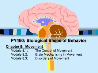
PY460: Biological Bases of Behavior. Chapter 8: Movement Module 8.1: The Control of Movement Module 8.2: Brain Mechanisms in Movement Module 8.3: Disorders of Movement. Slide 2 : The Control of Movement. Introduction: Clip #10: Sensory Motor Integration

E N D
PY460: Biological Bases of Behavior • Chapter 8: Movement • Module 8.1: The Control of Movement • Module 8.2: Brain Mechanisms in Movement • Module 8.3: Disorders of Movement
Slide 2: The Control of Movement Introduction: Clip #10: Sensory Motor Integration • movement-- an extremely complex process • complex “motor control” often w/o thought • Muscles-- “The Final Path”- multiple fibers • Smooth Muscle • movement of internal organs • stomach, arterial lining • Cardiac muscles (myocardium)- • interconnected bands of muscle • Skeletal Muscles- striated • long cylindrical fibers- “striped appearance”
Slide 3: Muscle Movement: Axons and Acetylcholine • Axon to fiber ratio- greater the ratio the more precise the movements [class; “typing with & w/o mittens] • e.g., eye= 1:3 arm (bicep) 1:100 • Neuromuscular junction- where the “motor neuron” meets a muscle fiber • NTR of movement- acetylcholine • effect: “contraction”, no Ach = relaxation • Myasthenia Gravis- An “autoimmune disease”- body attacks acetylcholine receptors • 2-3 per 100,000 over 75 years of age • Symptoms-progressive weakening and rapid fatigue of striated muscles as receptors are gradually destroyed. • Treatment-Immune suppressants & drugs inhibiting Acetylcholinesterase
Slide 4: Muscles Types and Functions • Antagonistic Muscles- opposing sets of muscles • Flexors- flexes or raises muscles • Extensors- extends or straightens • Fish Muscles- (movements & duration) • red (slow & long), pink (slow & not as long), white (fast & short) • Chicken Muscles (“white and dark meat”) • breast- fast acceleration, short duration • leg- long duration, not as fast (walking). • Human Muscles • Fast Twitch (anaerobic) • sprints/fast acceleration • Slow Twitch (aerobic) • duration/slow acceleration, speed
Slide 5: Proprioceptors-Feedback on Position & Movement • Proprioceptor: a receptor on the muscle sensitive to changes in muscle position and movement (“stretch”) of muscle. Respond with muscle contraction • Stretch Reflex- mediated at the spinal cord level • Muscle Spindle- stretch receptor attached parallel to muscle fibers sensitive to elongation of fibers • knee-jerk response • Golgi Tendon Organ- responds to increases in muscle tension. • Prevents excessive vigorous contraction (which would occur without it) • Life with reduced proprioception (babies, case in text)
Slide 6: Voluntary/Involutary Movements & Feedback • Types of Movements • Ballistic- large reflexive (all or none type) movements • Few ballistic movements-- most subject to feedback modifications Limits on Voluntary and Involuntary movement • few strictly involuntary, few strictly voluntary • limits of each (try swallowing 10 times) • Parkinson patients walking characteristics • INFANT REFLEXES: grasp reflex, Babinski reflex, rooting reflex, allied reflex • presence in adults signal damage to Cerebral Cortex
Slide 7: Coordinated Movement • Central Pattern Generator- proposed mechanism in spinal cord or brain that generates rhythmic patterns of “coordinated motor activity” that is extreme regular within species • stimulation of this mechanism affect action, but not frequency (apparently) • dog shaking off water, scratching reflex • Sequence of movements (e.g., walking) called a “Motor Program” • can be learned and built in. Think of a few! • Can be part of evolutionary inheritance • Yawning
Slide 8: The Spinal Cord-- Motor Program Keeper • How is it that a chicken can run without its head? • In humans • chewing, swallowing, breathing are controlled below the brain at the level of spinal cord/medulla. • Some motor programs (scratch reflex) are independent of brain feedback altogether • isolating of “scratch reflex” neurons from brain axons does not affect intrinsic firing rate and subsequent behavior. • Rhythm of firing even unaffected by muscular paralysis (the neurons are autorhythmic)
Slide 9: Brain and Movement (Begin Module 2) • Areas to be discussed • Cortical Areas in Movement • Primary Motor Cortex- messages (axons) to the medulla and spinal cord (just anterior to the precentral gyrus of the cerebral cortex) • control of “complex movement plans” • not reflexive (sneezing, cough, gag, cry etc.) • Areas near Primary Motor Cortex • Medulla and Spinal Cord- receive messages from PMC, control muscle movements (reflexive, bilateral, peripheral) • not much in chap 8, but see table 8.1 • Basal Ganglia & Cerebellummoderate movements but do not directly cause. (“selection, order, smoothing & future precision”)
Slide 10: The Cerebral Cortex • Primary Motor Cortex • Fritsch & Hitzig- ESB of PMC= coordinated movement • No direct connections to muscles, rather controls “complex movement plans” involving several muscles, not individual muscles. • i.e., activates central pattern generators • see fig 8.9- “motor homunculus” • See Figure 8.10- distribution of cells activated during hand movements
Slide 11: Working with the Primary Motor Cortex- Adjacent Areas • Posterior Parietal Cortex- control actions related to visual or somatosensory stimuli. • “cannot walk toward something they see” • Prefrontal Cortex- active in planning a potential movement- responds to sensory stimuli (future movement planning) • Premotor Cortex- active in preparation for movement, not during movement though. • Supplementary Motor Cortex- active during planning stage for rapid series of movements that require starting one movement before finishing another • e.g., Typing • Preparation for Movement- • coordinated waves of activity among these structure sending complex signals to PMC then down to the medulla and spinal cord.
Slide 12: Brain to Spinal Cord: 2 Tracks of Action • Dorsolateral Tract- axons projecting from PMC and Red Nucleus of Midbrain • axons cross over to opposite side of body controlling peripheral unlearned fine movements. • hands, fingers, toes • sometimes called the pyramidal tract • Ventromedial Tract- axons from PMC and SMC • axons branch to both sides- damage affects coordinate “side to side movements” like walking, standing, sitting, “twisting”, that is “bilateral movements”. • Neck, shoulders, trunk • 2 tracks act together to produce complete set of function muscle movements
Slide 13: The Cerebellum- “Follow My Finger” • Cerebellum- important in learned motor responses • programs allowing rapid sequential movement • damage-- trouble with rapid motor sequences requiring accuracy and timing • tapping to a rhythm • speaking • “adapting to prisms that distort vision” • “Saccades”- ballistic eye movements from one fixation point to another • damage or drunkenness (cerebellum 1st place affected by drink)- many small movements to fixate • Finger-to-Nose- inaccurate first movement, finger wavers during “hold”
Slide 14: Cellular Organization of Cerebellum& Duration of Movement • Perpendicular Organization of Cerebellar Cortex • precisely organized cellular structure • Parallel Fibers • Purkinge Cells (transmit to interior) • fire separately • inhibitory • Duration of movement • affected by number of Purkinge cells affected by parallel fiber excitation
Slide 15: Basal Ganglia: Organizing Planned Movements • Basal Ganglia has many roles- damage often results in much more than movement problems (e.g., memory, problem solving). • but some insight on its contributions to movement • seems to help in organizing new and habitual movements and inhibit unwanted movements (caudate nucleus) • e.g., signing your name • study of clumsy children
Slide 16: Parkinson’s Disease [video] • Symptoms- gradually increasing muscles tremors, slowed movement, inaccurate aim, difficult initiating physical, mental activity • Muhammad Ali • Prevalence: 1 per 100 over age 50 • Physiology- cell degradation in the substantia nigra & amygdala • decreased dopamine at D1 and D2 receptors resulting in net inhibitory response, thus “downstream” decreased excitation by cerebral cortex and thalamus • Natural degradation with age, some start with less cells, or lose at faster rate than others. • [Early and Late-Onset Parkinson’s (p.243)]
Slide 17: Etiology and Treatment of Parkinson’s • Suggested Causes • Inheritance • Interrupted blood flow to areas of the brain • Previous encephalitis or viral infection • Prolonged exposure to drugs/toxins • unlikely however that cause of most cases are due to drug abuse or exposure to toxin (Paraquat) (MPP+ MPTP). • Likely these factors contribute to process of degradation already active • Treatment: L-Dopa-cross BBB converted to dopamine. • Stereotyped movements, Delusions, Hallucinations • A “window” where helpful, soon disease too severe • Nicotine-- Smoking?? • Other Therapies [p.245]
Slide 18: Huntingdon’s Disease • A severe neurological disorder marked by gradually worsening tremors/twitches to severe writhing affecting daily movements like talking, walking, eating etc. • Prevalence: 1 per 10,000 • Widespread brain damage, particular area releasing GABA an inhibitory neurotransmitter • especially in basal ganglia (caudate nucleus etc.) • Genetic Conditions/Considerations • A dominant mutant gene.. Thus parent has 50% chance of passing disorder on. • Can test for the gene to determine not only who will get, but approximately when. • In vitro testing, other ethical issues
Slide 20: Spinal Cord Disorders • Paralysis- • Paraplegia- • Quadriplegia- • Poliomyelitis- • Lou Gehrig’s Disease- • Others • Flaccid Paralysis • Spastic Paralysis • Tabes Dorsalis