Download
1 / 78
840 likes | 1.34k Views
Diseases of White Blood Cells. Lecturer :Yiran Ni, MD Department of Pathology China Three Gorges University Email:nyr1986@gmail.com. Dec.2012. categories.

E N D
Diseases of White Blood Cells Lecturer :Yiran Ni, MD Department of Pathology China Three Gorges University Email:nyr1986@gmail.com Dec.2012
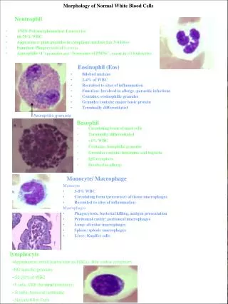
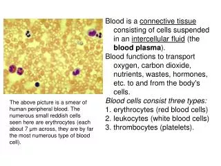
categories Disorders of white blood cells can be classified into two broad categories: proliferativedisorders, in which there is an expansion of leukocytes, and leukopenias, which are defined as a deficiency of leukocytes.
categories • Proliferations of white cells can be reactive or neoplastic. • Since the major function of leukocytes is host defense, reactive proliferation in response to an underlying primary, often microbial, disease is fairly common. Neoplastic disorders, although less frequent, are much more important clinically.
In the following lessons, we will first describe the 1\leukopenic states and summarize the common 2\reactive disorders and then consider in some detail 3\malignant proliferations of white cells.
Target of this class • Leukopenia; • Reactive leukocytosis; • Lymphadenitis; • General aspects and classification of neoplastic proliferation of white cell; • Acute myeloid leukaemia
Target of the next 2 class • chronic myeloid leukaemia • Lymphoma/lymphoid leukaemia • splenomegaly • histiocytoses • Review the slices we have learned
Leukopenia • The number of circulating white cells may be markedly decreased in a variety of disorders. • An abnormally low white cell count (leukopenia) usually results from reduced numbers of neutrophils (neutropenia, granulocytopenia).
Leukopenia • Lymphopenia is less common; in addition to congenital immunodeficiency diseases, it is most commonly observed in specific settings, such as advanced HIV infection, following therapy with glucocorticoids or cytotoxic drugs, autoimmune disorders, malnutrition, and certain acute viral infections. • Only the more common leukopenias involving granulocytes will be discussed further here.
Pathogenesis. • A reduction in circulating granulocytes will occur if there is (1) reduced or ineffective production of neutrophils or (2) accelerated removal of neutrophils from the circulating blood.
Pathogenesis. • (1) Inadequate or ineffective granulopoiesis is observed in the setting of: • 1. Suppression of myeloid stem cells, as occurs in aplastic anemia and a variety of infiltrative marrow disorders (tumors, granulomatous disease, etc.); • in these conditions, granulocytopenia is accompanied by anemia and thrombocytopenia.
Pathogenesis. • 2. Suppression of committed granulocytic precursors due to exposure to certain drugs. • Drugs are responsible for most of the significant neutropenias . Certain drugs, such as alkylating agents and antimetabolites used in cancer treatment, produce agranulocytosis in a predictable, dose-related fashion. • Because such drugs cause a generalized suppression of the bone marrow, production of erythrocytes and platelets is also affected
Pathogenesis. • 3. Disease states associated with ineffective granulopoiesis, such as megaloblastic anemias due to vitamin B12 or folate deficiency and myelodysplastic syndromes, where defective precursors are susceptible to death in the marrow. • 4. Rare inherited conditions (such as Kostmann syndrome) in which genetic defects in specific genes result in impaired granulocytic differentiation.
Pathogenesis. • (2) Acceleratedremoval or destruction of neutrophils occurs with • 1. Immunologically mediated injury to the neutrophils, which may be idiopathic, associated with a well-defined immunologic disorder (e.g., systemic lupus erythematosus), or produced by exposure to drugs.
Pathogenesis. • 2. Splenic sequestration, in which excessive destruction occurs secondary to enlargement of the spleen, usually associated with increased destruction of red cells and platelets as well. • 3. Increased peripheral utilization, as may occur in overwhelming bacterial, fungal, or rickettsial infections.
Morphology • Bone marrow: • The anatomic alterations in the bone marrow vary according to the underlying cause. • When neutropenia is caused by excessive destruction of mature neutrophils, the marrow is usually hypercellular owing to the presence of increased numbers of granulocytic precursors.
Morphology Bone marrow: • Hypercellularity is also the rule in neutropenias associated with ineffective granulopoiesis, as occurs in megaloblastic anemias and myelodysplastic syndromes. • Agranulocytosis caused by agents that suppress or destroy granulocytic precursors is understandably associated with marrow hypocellularity.
Morphology Other organ: • Infections (most often bacterial or fungal) are a common consequence of agranulocytosis. • Ulcerating necrotizing lesions of the gingiva, floor of the mouth, buccal mucosa, pharynx, or anywhere within the oral cavity are quite characteristic. • These ulcers are typically deep, undermined, and covered by gray to green-black necrotic membranes from which numerous bacteria or fungi can be isolated. • Less frequently, similar ulcerative lesions occur in the skin, vagina, anus, or gastrointestinal tract.
Morphology Other organ: • Severe life-threatening invasive bacterial or fungal infections can occur in the lungs, urinary tract, and kidneys. The neutropenic patient is at particularly high risk for deep fungal infections caused by organisms such as Candida and Aspergillus. • Sites of infection often show a massive growth of organisms with little leukocytic response. In the most dramatic instances, bacteria grow in colonies resembling those seen on nutrient media. • The regional lymph nodes draining these infections are enlarged and inflamed.
Clinical Course • The symptoms and signs of neutropenias are related to bacterial or fungal infections. They include malaise, chills, and fever, followed in sequence by marked weakness and fatigability. • In severe agranulocytosis with virtual absence of neutrophils, these infections can be overwhelming and cause death within a few days.
Clinical Course • A neutrophil count of less than 1000 cells per mm3of blood is worrisome, but most serious infections occur with counts below 500 per mm3. • Because infections are often fulminant, broad-spectrum antibiotics are given expeditiously whenever signs or symptoms appear.
Clinical Course • In some instances, such as following myelosuppressive chemotherapy, neutropenia is treated with granulocyte colony-stimulating factor (G-CSF), a growth factor that stimulates the production of granulocytes from marrow precursors
Reactive Proliferations of White Cells • Definition: Leukocytosis refers to an increase in the number of blood leukocytes. • It is a common reaction to a variety of inflammatory states and is sometimes the first indication of neoplastic growth of leukocytes
Pathogenesis • The peripheral blood leukocyte count is influenced by several factors, including: • 1. The size of the myeloid (for granulocytes and monocytes) and lymphoid (for lymphocytes) precursor and storage cell pools in the bone marrow, circulation, and peripheral tissues.
Pathogenesis • 2. The rate of release of cells from the storage pool into the circulation • 3. The proportion of cells that are adherent to blood vessel walls at any time • 4. The rate of extravasation of cells from the blood into tissues
Pathogenesis • leukocyte homeostasis is maintained by cytokines, growth factors, and adhesion molecules through their effects on the commitment, proliferation, differentiation, and extravasation of leukocytes and their progenitors.
Pathogenesis • In acute infection, there is a rapid increase in the egress of mature granulocytes from the bone marrow pool. • The release of IL-1, TNF, and other inflammatory cytokines stimulates bone marrow stromal cells and T cells to produce increased amounts of colony-stimulating factors (CSFs), which enhance the proliferation and differentiation of committed granulocytic progenitors and, over several days, cause a sustained increase in neutrophil production
Pathogenesis • Other growth factors preferentially stimulate other types of leukocytosis. • For example, IL-5 causes eosinophilia by enhancing the growth, survival, and differentiation of eosinophils, while IL-7 plays a central role in lymphopoiesis. • Such factors are differentially produced in response to various pathogenic stimuli
Pathogenesis • In most instances, it is not difficult to distinguish reactive leukocytosis from leukocytosis caused by flooding of the peripheral blood by neoplastic white blood cells (leukemia). • Uncertainties may arise in two settings: • 1. Particularly in children, acute viral infections can produce the appearance of activated lymphocytes in the peripheral blood and marrow that resemble neoplastic lymphoid cells.
Pathogenesis • 2. At other times, particularly in inflammatory states and severe chronic infections, many immature granulocytes appear in the blood, simulating a picture of myelogenous leukemia (leukemoid reaction). • Special laboratory studies (discussed later) are helpful in distinguishing reactive and neoplastic leukocytoses.
Reactive Proliferations of Lymph Nodes • In addition to causing leukocytosis, infections and inflammatory stimuli often elicit immune reactions within lymph nodes. • The infections that lead to lymphadenitis are numerous. • Most cause stereotypic patterns of lymph node reaction designated acute and chronic nonspecific lymphadenitis
Acute nonspecific lymphadenitis • Lymph nodes undergo reactive changes whenever they are challenged by microbiologic agents, cell debris, or foreign matter introduced into wounds or into the circulation. Acute lymphadenitis is most often seen: • 1. in the cervical region due to microbial drainage from infections of the teeth or tonsils
Acute nonspecific lymphadenitis • 2. in the axillary or inguinal regions secondary to infections in the extremities • 3. mesenteric lymph nodes draining acute appendicitis. • 4. Systemic viral infections (particularly in children) and bacteremia often produce generalized lymphadenopathy.
Morphology • Macroscopically, the nodes become swollen, gray-red, and engorged. • Histologically, there is prominence of the lymphoid follicles, with large germinal centers containing numerous mitotic figures. • Macrophages often contain particulate debris of bacterial origin or derived from necrotic cells.
Morphology • When pyogenic organisms are the cause of the reaction, the centers of the follicles may undergo necrosis, the entire node can sometimes be converted into a suppurative mass. • With less severe reactions, there is a neutrophilic infiltrate about the follicles, and numerous neutrophils can be found within the lymphoid sinuses. The cells lining the sinuses become hypertrophied and cuboidal and often undergo hyperplasia.
Clinical appearence • nodes with acute lymphadenitis are enlarged because of the cellular infiltration and edema. • As a consequence of the distention of the capsule, they are tender to touch. When abscess formation is extensive, they become fluctuant. • The overlying skin is frequently red, and sometimes penetration of the infection to the skin surface produces draining sinuses, particularly when the nodes have undergone suppurative necrosis. As might be expected, healing of such lesions is associated with scarring.
Chronic nonspecific lymphadenitis • lymph nodes in chronic reactions are not tender, because their capsules are not under increased tension. • Chronic lymphadenitis is particularly common in inguinal and axillary nodes, which drain relatively large areas of the body and are frequently challenged.
Neoplastic Proliferations of White Cells • Malignant proliferative diseases constitute the most important disorders of white cells. • These diseases can be classified into several categories: • 1. Lymphoid neoplasms • 2. Myeloid neoplasms • 3. histiocytoses
Categories • Myeloid neoplasmsarise from hematopoietic stem cells that give rise to cells of the myeloid (i.e., erythroid, granulocytic, and/or thrombocytic) lineage. Three categories of myeloid neoplasia are recognized: 1. acute myelogenous leukemias, in which immature progenitor cells accumulate in the bone marrow; 2. myelodysplastic syndromes, which are associated with ineffective hematopoiesis and resultant peripheral blood cytopenias; and 3. chronic myeloproliferative disorders, in which increased production of one or more terminally differentiated myeloid elements usually leads to elevated peripheral blood counts.
Categories • Lymphoid neoplasmsencompass a diverse group of entities. In many but not all instances, the phenotype of the neoplastic cell closely resembles that of a particular stage of normal lymphocyte differentiation, a feature that is used in the diagnosis and classification of these disorders.
Categories • The histiocytosesare uncommon proliferative lesions of macrophages and dendritic cells. • "histiocyte" is often applied to cells of macrophage or dendritic-cell lineage. • A special category of immature dendritic cells referred to as Langerhans cells gives rise to a spectrum of neoplastic disorders, some of which behave as disseminated malignant tumors, and others as localized benign proliferations. This group is called Langerhans cell histiocytoses.
Etiology and pathogenesis • Chromosomal translocations and oncogenes. Nonrandom chromosomal abnormalities, most commonly translocations, are present in the majority of white cell neoplasms. Many specific rearrangements are associated with particular neoplasms, suggesting a critical role in their genesis.
Etiology and pathogenesis • Chromosomal translocations frequently occur in myeloid neoplasms
Etiology and pathogenesis • 2. Inherited genetic factors:individuals with genetic diseases that promote genomic instability. • telangiectasia, are at increased risk for development of acute leukemia. In addition, both Down syndrome (trisomy 21) and neurofibromatosis type I are associated with an increased incidence of childhood leukemia.
Etiology and pathogenesis 3. Viruses. Three viruses-human T-cell leukemia virus-1 (HTLV-1), Epstein-Barr virus (EBV), and Kaposi sarcoma herpesvirus/human herpesvirus-8 (KSHV/HHV-8) have been implicated as causative agents.
Etiology and pathogenesis • HTLV-1 has been associated only with adult T-cell leukemia/lymphoma. • EBV genomes are found in the tumor cells of a subset of Burkitt lymphoma, 30% to 40% of cases of Hodgkin lymphoma, many B-cell lymphomas occurring in the setting of T-cell immunodeficiency, and rare natural killer cell lymphomas. • KSHV is uniquely associated with an unusual type of B-cell lymphoma that presents as a malignant effusion, often in the pleural cavity.
Etiology and pathogenesis • 4. Environmental agents. • The most clear-cut associations are those of Helicobacter pylori infection with gastric B-cell lymphoma and gluten-sensitive enteropathy with intestinal T-cell lymphoma.
Etiology and pathogenesis • 5. Iatrogenic factors. • Radiotherapy and certain forms of chemotherapy used to treat cancer increase the risk of subsequent myeloid and lymphoid neoplasms. • This association is believed to stem from mutagenic effects of ionizing radiation and chemotherapeutic drugs on hematolymphoid progenitor cells.
Myeloid neoplasm • The common feature that unites this heterogeneous group of neoplasms is an origin from hematopoietic progenitor cells capable of giving rise to terminally differentiated cells of the myeloid series (erythrocytes, granulocytes, monocytes, and platelets).