Download
1 / 43
430 likes | 616 Views
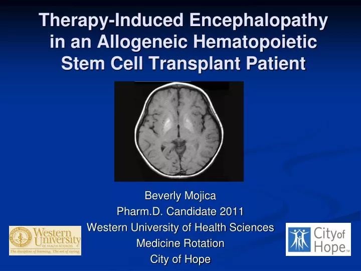
Therapy-Induced Encephalopathy in an Allogeneic Hematopoietic Stem Cell Transplant Patient. Beverly Mojica Pharm.D. Candidate 2011 Western University of Health Sciences Medicine Rotation City of Hope. Outline. Patient case Background of encephalopathy

E N D
Therapy-Induced Encephalopathy in an Allogeneic Hematopoietic Stem Cell Transplant Patient Beverly Mojica Pharm.D. Candidate 2011 Western University of Health Sciences Medicine Rotation City of Hope
Outline Patient case Background of encephalopathy Causes of encephalopathy in HSCT recipients Drug-induced encephalopathy Tacrolimus Methotrexate Cytarabine Rituximab Future directions Questions
Case: N.B. N.B. is a 62 year-old male (186.2 cm, 70.7 kg) with a history of mantle cell lymphoma with CNS involvement Admitted on 5/12/10 to City of Hope for an allogeneic stem cell transplant from a matched unrelated donor
Case: N.B. Past Medical History: History of prostate cancer Refractory mantle cell lymphoma with CNS involvement (leptomeningeal) Bell’s Palsy Family History: Father had prostate cancer Sister had an aneurysm and lives in Holland Social History: Supported by his family including his wife Engineer who is self-employed as a consultant in the Siemens/Diagnostic Imaging machines Smoked 1 pack of cigarettes for 20 years (quit in 1979) Drinks alcohol occasionally
Case: N.B. Pertinent Medications:
Case: N.B. • Clinical History • 5/27: • Confused overnight • Tmax: 37 C • 5/28: • Hallucinations in the morning • Tmax: 37.2 C • 5/29: • Confused overnight • Blank staring • Keppra 500 mg BID • Tmax: 37.1 C
Case: N.B. • Microbiology • 6/4: • Stool: rapid CMV and HSV shell vial cultures negative • 6/5: • Aspergillus Ag negative by EIA • Cryptococcal Ag serum negative • Fungitell 1,3 Beta D glucan negative (65 pg/mL) • Toxoplasma gondii from serum not detected
Case: N.B. • Electroencephalogram (EEG) • Slow background (6 Hz) consistent with encephalopathy • MRI Head • 6/9: No mass effect or focal abnormality • Cytology (spinal tap from omaya catheter) • 5/20 : No lymphoma in CSF • 6/15 : No lymphoma in CSF
Encephalopathy1 Any diffuse disease of the brain that alters brain function or structure Causes: Infection (bacteria, virus, or prion) Metabolic or mitochondrial dysfunction Brain tumor or increased intracranial pressure Exposure to toxins (i.e. solvents, drugs, alcohol, paints, industrial chemicals, and certain metals) Radiation Trauma Poor nutrition Ischemia
Causes of Encephalopathy in Allogeneic HSCT Recipients2 • Infection • Fungi (Aspergillus, Candida), Gram-positive bacteria Toxoplasma organisms, Viral (CMV, human herpes virus 6 or 7, Epstein-Barr, varicella-zoster) • Vascular Disorders • Thrombocytopenia, thrombosis, embolism • Tumor • Lymphoproliferative Disorders • Therapy-related encephalopathy
Tacrolimus3,4 MOA: potent inhibition on T-lymphocyte activation by inhibiting calcineurin phosphatase activity
Tacrolimus3,6 • Absorption: Oral: Incomplete and variable • Distribution: 0.55-2.47 L/kg • Metabolism: Extensively hepatic via CYP3A4 to eight possible metabolites • Excretion: • Feces (~93%) • Urine (<2% as unchanged drug) • Biological half-life varies: 3.5-40.5 hours3
Tacrolimus Neurotoxicity • Incidence: ~5-30%7 • Posterior reversible encephalopathy syndrome (PRES) • Initial manifestation: • Sudden altered mental status, confusion, headache, diminished spontaneity and speech, lethargy, unconsciousness, convulsions • Not dose-dependent2,6,7,8 • Can occur at anytime after HSCT (usually within 1 month)
Tacrolimus Neurotoxicity2,5,6 • Mechanism unclear: • Direct endothelial damage injury to the capillary bed alteration of blood-brain barrier (BBB) white matter edema release of vasoactive peptides (endothelin, thromboxane, prostacyclin) vasospasm or interruption of cerebral autoregulation
Tacrolimus Neurotoxicity5,7 • Radiologic Findings • MRI: edema involving white matter in the posterior portions of the cerebral hemispheres (esp. bilaterally in the parieto-occipital regions); hyperintense lesions (T2 weighted) • CT: low attenuation of white matter • EEG: diffuse slowing or sharp epileptic discharges
Methotrexate9,10 MOA: inhibits DNA synthesis by irreversibly binding to dihydrofolate reductase
Methotrexate10,11 • Absorption: completely absorbed with parenteral route • Distribution: widely distributed throughout body • Metabolism: <10% with hepatic aldehyde and intestinal bacteriaoxidase • Excretion: renal (~90% unchanged in the urine); small amount in feces • Renal impairment: CNS half-life may reach 19-44 hours • Half-life: 4.5 -14 hours
Methotrexate Neurotoxicity12,13,14 • Acute • Onset: during or within hours after MTX • Somnolence, confusion, fatigue, seizures • Usually reversible • Subacute (3-15%) • Onset: days to weeks post MTX treatment • Stroke-like syndrome • Hemiparesis, seizures, speech disorder • Usually reversible • Chronic • Onset: months to years • Leukoencephalopathy • Dementia, focal seizures, quadriparesis, stupor • May or may not be reversible
Methotrexate Neurotoxicity15,16 • Incidence2 • < 10% with high dose IV MTX2 • Up to 40% with IT2 • Risk factors: • Dose-related • Age >10 • Cranial irradiation • Concomitant use of cytarabine, daunorubicin, salicylates, sulfonamides or vinca alkaloids
Methotrexate Neurotoxicity12,14,15,17,18 • Mechanism not well established • Direct toxic effects on neurons • MTX inhibits dihydrofolate reductase • Increased levels of adenosine • Dilation of cerebral blood vessels • Decreased synthesis of biogenic amine neurotransmitters • Elevated homocysteine: • Endothelial cell injury • Cerebrovascular infarcts
Methotrexate Neurotoxicity • Route (IV, IT) and dose-dependent (cumulative exposure) • IV > 1 g/m2 (or frequent IV)12,16,18 • IT: 12-15 mg (> 100 mg)19 • Higher risk when IT MTX >50 mg in combination with cranial irradiation or systemic (IV) MTX 15 • Recurrence rate: 10-56% upon rechallenge18 • IT MTX must be preservative-free18
Methotrexate Neurotoxicity14,15 • Management • Antidote for reversal of MTX neurotoxicity: aminophylline 2-5 mg/kg every 6 hours14,15,18 • Displaces adenosine from the receptor • IT MTX overdose: glucarpidase 50 units/kg bolus IV injection over 5 minutes 10 • Rapidly decrease MTX levels by up to 98% in 30 minutes • Not available commercially • Call: 1-866-918-1731 for overnight shipping
Methotrexate Neurotoxicity10,16 • Prevention • Folinic acid (leucovorin rescue) • 100 mg/m2 48 hours after MTX administration q 3 hours x8 doses followed by 200 mg/m2 q 6 hours x4 doses16 • High dose did not compromise cure16 • Hydration10 • 2.5 -3.5 L/m2 per day starting 12 hours prior to MTX infusion • Urinary alkalinazation10 • 50 mL of D5W containing sodium bicarbonate 1 mEq/kg IV over 30 minutes q 4-6 hours
Cytarabine20 • MOA: primary action is inhibition of DNA polymerase resulting in decreased DNA synthesis and repair. • Cytarabine is specific for the S phase of the cell cycle (blocks progression from the G1 to the S phase).
Cytarabine20,21,22 • Absorption: Complete with IV • Distribution: Widely and rapidly in most tissues • Crosses BBB with CSF levels of 40% to 50% of plasma level • Metabolism: Primarily hepatic; 86% to 96% of dose is metabolized to inactive metabolite • IT little conversion to inactive metabolite • Excretion: Renal (~80%; 90% as inactive metabolite) within 24 hours • Half-life • IV: < 20 minutes21 • IT: 2-6 hours 20,21
Cytarabine Neurotoxicity13,21,23 • Route: Intrathecal, IV, liposomal23,13 • Cerebellar dysfunction (most common), generalized encephalopathy, peripheral neuropathy, and arachnoiditis, fecal and urinary incontinence • Cytotoxic levels of cytarabine may be maintained for up to 24 hours after IT administration • IT liposomal (sustained release) may maintain cytotoxic concentrations of the drug in the CSF for up to 14 days • CSF exposure up to 40x that of standard Ara-C • Onset: As early as 2-5 days after treatment13 • May resolve spontaneously within a few days or may be permanent23,13
Cytarabine Neurotoxicity13,23 • Incidence: varies from 5-50%13,24 • Risk factors13,23 • IV doses > 1 g/m2 23 • Total IV dose > 30 g (> 3g/ m2 every 12 hours)3 • IT dose (> 100 mg per week)21 • Age > 40 years of age13 • Prior cytarabine therapy • Renal dysfunction • IT, IT liposomal administration13 • Concomitant use with high-dose chemotherapy (i.e. methotrexate)13,24
Cytarabine Neurotoxicity25,26 • MOA of how it causes encephalopathy: -Cytotoxic effect25 -Immune-mediated mechanism is hypothesized26 • Management: • Cytarabine should be discontinued immediately13 • No standard treatment is available • Corticosteroids (methylprednisolone, dexamethasone)25,26 • Prevention • Concurrent use of corticosteroid with IT liposomal cytarabine reduces risk of arachnoiditis21,24,25
Rituximab27 • MOA: B cell lysis by binding of the Fab domain of rituximab to the CD20 antigen on B lymphocytes and by recruitment of immune effector functions by the Fc domain • Complement-dependent cytotoxicity (CDC) • Antibody-dependent cellular cytotoxicity (ADCC)
Rituximab27,28,29 • Absorption: I.V.: Immediate and results in a rapid and sustained depletion of circulating and tissue-based B cells • Metabolism: Hepatic • Distribution: Lymph nodes • Excretion: Uncertain; may undergo phagocytosis and catabolism in the reticuloendothelial system (RES) • Median terminal half-life for NHL: 22 days (range: 6-52 days)
Rituximab Neurotoxicity30 • Progressive Multifocal Leukoencephalopathy (PML) • Incidence: Rare • 2 PML cases per 8000 rituximab treated SLE patients • Need to conduct more epidemiological studies • Risk factors: • Need more studies • Possibly low CD4 counts and low IgG levels
Rituximab Neurotoxicity27,30 • Clinical presentation • Confusion/disorientation • Motor weakness/hemiparesis • Altered vision/speech • Poor motor coordination • Symptoms progress over weeks to months • MOA of how it causes encephalopathy1,2 • Unclear, but rituximab can decrease the immune system and cause reactivation of the Jakob-Creuzfeld (JC) virus
Rituximab Neurotoxicity30 • A retrospective analysis of patients diagnosed with PML after rituximab treatment • Cases from cancer centers or academic hospitals (22), FDA reports (11), manufacturers database (30), publications (18) • Inclusion: rituximab therapy prior to PML, PML confirmation with brain histology or MRI, no HIV infection • Patient Population (n=57) • B-cell lymphoproliferative disorder (52) • Systemic Lupus Erythmetous (2) • Autoimmune pancytopenia (2) • Immune thrombocytopenia purpura (1)
Rituximab Neurotoxicity30 • Onset: • Median of 16 months (following rituximab initiation) • 5.5 months (following last rituximab dose) • 6 rituximab doses preceded PML diagnosis • In the absence of immune reconstitution, case fatality rate was 90% • Survival rates up to 38% after hematopoietic stem cell transplantation
Rituximab Neurotoxicity27,30 • Promptly evaluate any patient presenting with neurological changes • Consider neurology consultation, brain MRI and lumbar puncture for suspected PM L • Discontinue rituximab in patients who develop PML • Consider reduction/discontinuation of concurrent chemotherapy or immunosuppressants • Risks versus benefits
Back to N.B. • Clinical History • 6/16: confusion is clinically improving • 6/17: mental status seems to be slowly improving • 6/20: confusion clinically stable • Acute altered mental status attributed to tacrolimus CNS toxicity
Future Directions • Need of biological markers or markers for quantification of medication-induced neurotoxicity • Adenosine • Choline (higher levels correlated with demyelination) • Patterns • MRI, CT, EEG
References 1. Author unknown. NINDS Encephalopathy Information Page. National Institute of Neurological Disorders and Stroke. http://www.ninds.nih.gov/disorders/encephalopathy/ encephalopathy.htm Last updated: 0212/2007. Date accessed: 06/17/2010 2. Nishiguchi T, Mochizuki K, Shakudo M, et al. CNS complications of Hematopoietic Stem Cell Transplantation. AJR 2009; 192: 1002-1011 3. Prograf® (tacrolimus) injection package insert. Astellas Pharma.Deerfield, IL. Last Revised: August 2009 5. Hinchey J, Chaves C, Appignani B, et al. A Reversible Posterior Leukoencephalopathy Syndrome. NJEM (1996) 334:494-500. 6.Oliverio P, Restrepo L, Mitchell S, et al. Reversible Tacrolimus-induced Neurotoxicity Isolated to the Brain Stem. Am J Neuroradiol (2000) 21: 1252-1254 7. Grimbert P., Azema C., Pastural M., et al. Tacrolimus (FK506)-induced severe and late encephalopathy in a renal transplant recipient. Nephrol Dial Transplant (1999) 14: 2489-2491. 8. Chegounchi M, Hanna M, Neild G. Progressive neurological disease induced by tacrolimus in a renal transplant recipient: Case presentation. BMC Nephrology 2006 Vol 7:1-3 9. Methotrexate for injection, USP package insert. Bedford Laboratories. Bedford, OH. Last Revised: April 2005.
References 10. LaCasce A. Therapeutic use of high-dose methotrexate. UpToDate. http://uptodate.com.proxy. westernu.edu/online/content/ topic.do?topicKey=chemge. Last updated: 02/23/2009 Date Accessed: 06/16/2010 11. Lexi-Comp online, Lexi-Drugs Online. Hudson, Ohio Lexi-Comp, Inc. Methotrexate. Last updated 6/18/10. Accessed 6/18/10. 12. Vazmar S, Schusseler P, Becker A, et al. Methotrexate-Associated Alterations of the Folate and Methyl-transfer Pathway in the CSF of ALL Patients with and without Symptoms of Neurotoxicity. Pediatr Blood Cancer (2009) 52:26-32 13. Wen P, Plotkin S. Neurologic complications on non-platinum cancer chemotherapy. UpToDate. http://www.uptodate.com.proxy.westernu.edu /online/contecnt/topic.do? topicKey=genl_onc Last updated: 12/27/2009. Date accessed: 06/17/2010. 14. Brugnoletti F, Morris EB, Laningham FH, et al. Recurrent Intrathecal Methotrexate Induced Neurotoxicity in an Adolescent with Acute Lymphoblastic Leukemia: Serial Clinical and Radiologic Findings. Pediatric Blood Cancer p.293-29515. 15. Shuper A, Stark B, Kornreich L, et al. Methotrexate-related Neurotoxicity in the Treatment of Childhood Acute Lymphoblastic Leukemia. IMAJ (2002) Vol 4 p1050-1051 16. Hamidah A, Lope R, Latiff Z, et al. Prevention of Neurotoxicity by High-dose Folinic Acid Rescue after High-dose methotrexate and Intrathecal methotrexate without Compromising Cure inspite of Previous Transient Leukoencephalopathy after Intrathecal Methotrexate. Annals Academy of Medicine. p743-744
References 17. Dicuonzo F, Salvati A, Palma M, et al. Posterior Reversible Encephalopathy Syndrome Associate with Methotrexate Neurotoxicity: Conventional Magnetic Resonance and Diffusion-Weighted Imaging Findings. J Child Neurol (2009) 24;8:1013-1018 18. Inaba H, Khan RB, Laningham FH, et al. Clinical and Radiological Characteristics of Methotrexate-induced Acute Encephalopathy in Pediatric Patients with Cancer. Annals of Oncology (2008) 19: 178-184 19. Finkelstein Y, Zevin S, Raikhlin-Eisenkraft B, Bentur Y. Intrathecal methotrexate neurotoxicity: clinical correlated and antidotal treatment. Environmental Toxicology and Pharmacology (2005) 19:721-725 20. Cytarabine for injection, USP package insert. Bedford Laboratories. Bedford, OH. Last Revised: September 2008 21. Kwong YL, Yeung D, Chan J. Intrathecal Chemotherapy for Hematologic Malignancies: Drugs and Toxicities. Ann Hematol (2009) 88: 193-201 22. Lexi-Comp online, Lexi-Drugs Online. Hudson, Ohio Lexi-Comp, Inc. Cytarabine. Last updated 4/7/2010. Accessed 6/17/10. 23. Nielsen E, Brant J. Chemotherapy-Induced Neurotoxicity. AJN Supplement (2002) 16-19
References 24. Jabbour E, O’Brien S, Kantarjian H, et al. Neurologic complications associated with intrathecal cytarabine given prophylatically in combination with high-dose methotrexate and cytarabine with acute lymphocytic leukemia. Blood (2007) Vol 109, No 8, pp 3214-3218 25. Hilgendorf I, Wolff D, Junghass C, et al. Neurological complications after Intrathecal liposomal cytarabine application in patients after allogeneic hematopoietic stem cell transplantation. Ann Hematol (2008) 87:1009-1012 26. Malhotra P, Mahi S, Lal V, et al. Cytarabine-Induced Neurotoxicity Responding to Methylprednisolone. American Journal of Hematology 77: 416 27. Rituxan® (rituximab) for injection package insert. Genentech, Inc. South San Francisco, CA. Las Revised: 02/2010 28. Lexi-Comp online, Lexi-Drugs Online. Hudson, Ohio Lexi-Comp, Inc. Rituximab. Last updated 6/18/10. Accessed 6/18/10. 29. Cartron G, Blasco H, Paintaud G, Watier H, Le Guellec C. Pharmacokinetics of rituximab and its clinical use: thought for the best use? Crit Rev Oncol Hematol 62(1):43-52 (2007 Apr). 30. Carson K, Evens AM, Richey EA. Progressive multifocal leukoencephalopathy after rituximab therapy in HIV-negative patients: a report of 57 cases from the Research on Adverse Drug Events and Reports project. BLOOD 05/14/2009. Vol 113, No 20. pp4833-4840.