1 / 5
50 likes | 65 Views
Since the first reports of laparoscopic surgery for inflammatory bowel disease from Peters in 1992, several literatures have subsequently shown the potential advantage of minimal access surgery for small bowel pathologies.

E N D
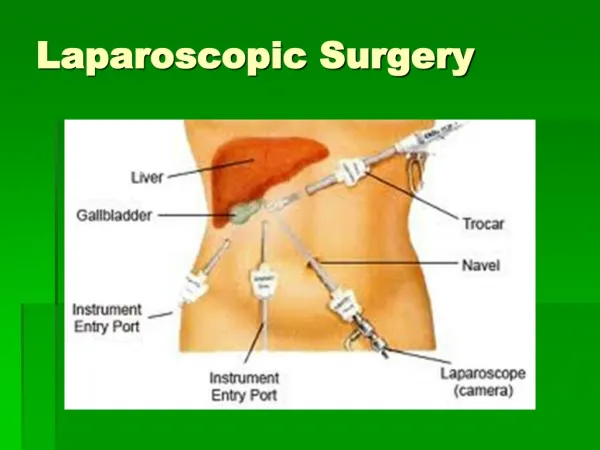
Laparoscopic Sm all Bo wel Surgery Prof. Dr. R. K. Mishra INTRODUCTION SURGICAL ANATOMY The bowel wall is comprised of four layers: (1) the mucosa, (2) submucosa, (3) muscularis propria, and (4) serosa. The submucosa is the “strength” layer of the anastomosis and must be included in every suture pass. The vascular supply to the bowel is via the mesentery. The superior mesenteric artery gives rise to the arcuate system that supplies the vasa recta of the small intestine. If an anastomosis is constructed off to one side of the bowel, there will be a shorter distance for the blood supply to reach the mesenteric aspect of the anastomosis. However, the blood supply to the antimesenteric aspect of the anastomosis must travel a considerably longer distance over the antimesenteric border of the bowel. That puts the antimesenteric aspect of the anastomosis at a much higher risk of ischemia. For this reason, we suggest centering side-to-side and side-to-end anastomoses on the antimesenteric border of the bowel, rather than off to one side, whenever possible, to minimize ischemia. Since the first reports of laparoscopic surgery for inflammatory bowel disease from Peters in 1992, several literatures have subsequently shown the potential advantage of minimal access surgery for small bowel pathologies. The increased use of laparoscopy in the management of gastrointestinal problems continues to expand. Procedures such as jejunostomies, diagnosis of intestinal obstruction or ischemia, resection of the small bowel, and lysis of adhesions can be managed with this technique. LAPAROSCOPIC RESECTION OF SMALL BOWEL The role of laparoscopy in resection of the small bowel has increased rapidly in the last 5 years. Laparoscopic small bowel resection can be performed safely in the setting of benign as well as malignant disease and imparts many of the benefits of minimally invasive surgery. The affected small bowel and mesentery can be thoroughly inspected and resected laparoscopically and bowel continuity restored through an entirely intracorporeal technique or laparoscopic-assisted technique. Staplers There are three standard types of surgical staplers used for bowel resection and reconstruction: 1. Transverse anastomosis (TA) staplers are typically noncutting straight staplers that lay down several rows of staples. After stapling with a TA stapler, the specimen needs to be transected with scissors or a scalpel. 2. Linear staplers, most commonly referred to as gastro- intestinal anastomosis (GIA) staplers, are similar to the TA staplers but have an additional cutting mechanism for transection. Three rows of staples are placed on either side of the cut. This type of stapler is ideal for laparoscopic bowel resection. 3. End-to-end anastomosis (EEA) staplers are circular cutting staplers that place several rows of staples on both cut ends as well. While the individual staples are almost always made of titanium, staplers come in a variety of configurations and lengths for open and laparoscopic procedures. Staplers can also accommodate different cartridges to handle different tissue thickness. Cartridges are preloaded with staples and Indications A small bowel resection is the surgical removal of one or more segments of the small intestine. Laparoscopic small bowel resection with primary anastomosis is most frequently indicated for benign diseases. The most common indications are: ■ Isolated Crohn’s disease ■ Gastrointestinal stromal tumors ■ Benign strictures, and ■ Vascular malformations Malignant conditions represent relative contra- indications in that they are rare, and if diagnosed or suspected, laparoscopic management should not be considered as the method of choice. The conduct of the laparoscopic operation should be in a manner very similar to that of a conventional small bowel resection.
335 CHAPTER25: Laparoscopic Small Bowel Surgery color coded by the manufacturer to indicate the height of the staples. Typical staple heights range from 2 mm for thin, vascular tissue (e.g., mesentery) to 4.1 mm for thick tissue (e.g., inflamed bowel/colon). The typical staple height used for the majority of bowel resection/anastomosis procedures is 3.5 mm. Linear Stapled Anastomosis Side-to-side stapled reconstructions start with sharply removing a portion of the staple line near the antimesenteric border if a stapler has been used to transect the bowel. This also allows bowel viability to be assessed; brisk bleeding from the cut edge implies adequate perfusion for the reconstruction. The GIA stapler is then introduced into each end of the bowel. Cartridge length is left to the surgeon’s discretion, but typically utilizes is a 60-mm load when performing a stapled anastomosis. Using one hand, the bowel is manipulated so that the antimesenteric borders of the bowel are included in the jaws of the stapler as they close. This helps ensure that the anastomosis is perfectly centered on the antimesenteric border of the bowel, which maximizes perfusion. The stapler is closed, and bowel inspected to ensure that no other intra-abdominal contents have been inadvertently caught by the stapler. Once this is confirmed, a reinforcing silk “crotch” stich may be placed at the distal end of the anastomosis as this area is subject to the most tension. The stapler is then fired. It is then removed and the newly created common channel is inspected for hemostasis with a ring forceps. Interrupted sutures can be used to control larger bleeders from the staple line, though most oozing will be self-limited. Nowadays, the braided absorbable suture is used to close the common enterotomy in an interrupted fashion. Full- thickness bites are used, taking care to invert the mucosa. The staple lines should be offset to minimize the potential ischemic area. Fig. 1: Position of the surgical team for ileal resection. Port Position Port should be positioned according to the baseball diamond concept and should be opposite to the site of pathology. For jejunal surgery, the left- and right-sided cannulae may suffice. For ileal surgery, it may be preferable to use the suprapubic cannula (Fig. 2). Operative Technique Once the preoperative diagnosis is confirmed and the surgeon is confident that laparoscopic procedure appears feasible, the pathology is located by walk over the entire length of the small intestine and placing a suture just upstream of the pathology. Walk over the small bowel is accomplished from proximal to distal by placing the patient on the left side up, in slight reverse Trendelenburg position until the mid-small bowel is reached, then adjusting the patient to the right side up with Trendelenburg position to run the distal half of the small intestine. The surgeon should start the “walk over” from between the legs then switch to the left side of the patient for the distal half. The technique of inspection should be “hand- over-hand” or “hand-to-hand” (Figs. 3A to D) based on the degree of freedom present within the abdominal cavity. If it is advantageous to divide the mesenteric vessels before delivery of the specimen through the abdominal incision, this should ideally be done using LigaSure (Fig. 4). Patient Positioning and Operating Room Setup The patient is placed supine in a modified lithotomy position using Dan Allen stirrups. Surgery is begun in the Trendelenburg position (20° head-down tilt) and, after cannula insertion, the patient is tilted left side down for ileal surgery or the right side down for jejunal surgery (Fig. 1). After insertion of the port, the surgeon stands between the legs. The scrub nurse should stand on the right side of the patient near the knee. The assistant stands on the right side of the surgeon, one monitor is placed close to the patient’s right shoulder, and the second monitor is situated near the left shoulder, the best location for viewing by the nurse.
336 SECTION2: Laparoscopic General Surgical Procedures The main vessel supplying the affected segment can also be ligated, leaving the other vessels of the mesentery to be divided through the incision. This may be especially helpful in a patient with a thick abdominal wall. Once the specimen is fully mobilized, a port site is enlarged to 3–5 cm. For small incisions, a transverse incision is preferred. The anterior rectus sheath is transversely incised, the rectus muscles retracted, and the posterior sheath also transversely incised. If the incision has to be larger because of a bulky tumor, a midline longitudinal incision in the midline is accomplished above and below the umbilicus. The wound is protected using a plastic sheath, and the loop of intestine to be resected is drawn out through the enlarged incision (Figs. 5A and B). Wound protection is important to reduce any contamination by tumor cells or intestine, and it may also facilitate the specimen extraction. The resection and anastomosis are then made in a standard manner extracorporeally, either by a hand-sewn or stapled method. The mesenteric defect is usually closed with a running absorbable suture through the incision (Figs. 6A and B). After performing the anastomosis, the abdomen is copiously irrigated with warm sterile saline solution through the incision. The fluid is removed by placing the patient in the head-up position and passing a sump suction cannula into the pelvis. After the irrigation of the peritoneal cavity, the abdominal wall is closed with a running suture or a single series suture. The most important steps of laparoscopically-assisted small bowel surgery are to localize and mobilize the diseased segment and deliver it through a small incision of hand port. The laparoscopic technique has become the procedure of choice for isolated benign small diseases. The intracorporeal Fig. 2: Position of the cannulae for ileal resection. A B C D Figs. 3A to D: Walk over to small intestine in order to detect pathology.
338 SECTION2: Laparoscopic General Surgical Procedures is also useful to evaluate periureteral inflammation and to aid in the decision to use intraoperative ureteric catheters. Preoperative enteral or parenteral nutritional support should be considered in selected patients. Most surgeons would agree that the laparoscopic approach is contraindicated in patients with nonlocalized intra-abdominal abscesses, multiple previous bowel operations with possible dense adhesions, fixed mass with multiple fistulas, acute intestinal obstruction, and perforation. Although the entire operation can be performed laparoscopically, most surgeons prefer a laparoscopic- assisted procedure by laparoscopic mobilization and extracorporeal resection and anastomosis. examined through the incision, and drain tube is left in cul- de-sac through the right lateral trocar site if necessary, and every trocar site incision is closed with skin staplers. Laparoscopic-assisted approach, with an extracorporeal anastomosis, rather than an entirely laparoscopic approach with an intracorporeal anastomosis is more popular. Laparoscopic-assisted approach provides the benefits of laparoscopic surgery while maintaining the advantages of open bowel division and anastomosis, i.e., speed and low risk of intra-abdominal stool spillage. Laparoscopic small bowel surgery for benign conditions continues to evolve. When performed by surgeons with adequate experience, laparoscopic surgery seems to demonstrate advantages over conventional operations. We believe at this time that laparoscopic ileocolectomy should be considered the first- line surgical option for most primary resections for Crohn’s disease localized to the ileocolic region. Operative Technique The patient is placed in the Trendelenburg position, and three or four ports are inserted according to baseball diamond concept. For establishment of pneumoperitoneum, CO2 is channeled through the infraumbilical trocar until the intra- abdominal pressure reaches 12 mm Hg. Both the operating surgeon and camera holder stand on the patient’s left side. After abdominal exploration, the operation table is rotated left side down so the small intestine falls toward the left upper quadrant. The ascending colon is thoroughly mobilized from the base of the appendix up to the hepatic flexure by cutting the retroperitoneal attachments with electrosurgical scissors and laparoscopic coagulating shears, and bluntly dissecting the retroperitoneal fusion fascia and loose connective tissue. With this procedure, the duodenum, Gerota’s fascia, and sometimes more inferiorly the right ureter and the gonadal vessels become visible beneath the retroperitoneal fusion fascia. During dissection, the direct grasping and handling of diseased bowel loops should be avoided, to prevent incidental myotomies and enterotomies. In patients of Crohn’s disease with ileovesical, ileorectal, and gastrocolic fistulas, division with an intracorporeal stapling device by one or two firings of the 45 or 60 mm stapler can be done. After mobilization of the entire ascending colon, meticulous hemostasis is ensured. Then, the patient is placed in a reversed Trendelenburg position temporarily and the abdomen is irrigated with warm sterile saline. Finally patient is placed in a flat supine position, and pneumoperitoneum is released. To exteriorize the bowel loop a small laparotomy is performed through a 5 cm long skin incision made at the umbilical trocar site or through a Pfannenstiel incision. A wound protector is inserted and the segments of the colon are delivered through this incision. Mesenteric division, ileocolic resection, and anastomosis by Gambee’s procedure using 4-0 absorbable sutures, or functional EEA using linear staplers are performed extracorporeally. After closure of the mesenteric defect, the entire residual small bowel is BIBLIOGRAPHY 1. Altman DG. Practical Statistics for Medical Research. London, England: Chapman and Hall; 1991. 2. American Thoracic Society. Standardization of spirometry: 1987 update. Am Rev Respir Dis. 1987;136:1285-98. 3. Azagra JS, Goergen M, Gilbart E, Jacobs D, Lejeune P, Carlier E. Anterior resection: The total laparoscopic approach. In: Monson JRT, Darzi A (Eds). Laparoscopic Colorectal Surgery. Oxford, England: Isis Medical Media; 1995. pp. 38-55. 4. Bartlett RH, Brennan ML, Gazzaniga AL, Hanson EL. Studies on the pathogenesis and prevention of postoperative pulmonary complications. Surg Gynecol Obstet. 1973;137:925-33. 5. Beecher HK. The measured effect of laparotomy on the respiration. J Clin Invest. 1933;12:639-50. 6. Benhamou D, Simonneau G, Poynard T, Goldman M, Chaput JC, Duroux P. Diaphragm function is not impaired by pneumoperitoneum after laparoscopy. Arch Surg. 1993;128:430-2. 7. Berstein MA, Dawson JW, Reissman P, Weiss EG, Nogueras JJ, Wexner SD. Is complete laparoscopic colectomy superior to laparoscopic assisted colectomy? Am Surg. 1996;62:507-11. 8. Böhm B, Nouchirvani K, Hucke HP, Stock W. Morbidity and mortality after elective resections of colorectal cancers [in German]. Langenbecks Arch Chir. 1991;376:93-101. 9. Bonnet F, Blery C, Zatan M, Simonet O, Brage D, Gaudy J. Effect of epidural morphine on post-operative pulmonary dysfunction. Acta Anaesthesiol Scand. 1984;28:147-51. 10. Chen HH, Wexner SD, Weiss EG, Nogueras JJ, Alabaz O, Iroatulam AJ, et al. Laparoscopic colectomy for benign colorectal disease is associated with a significant reduction in disability as compared with laparotomy. Surg Endosc. 1998;12:1397-400. 11. Christensen EF, Schultz P, Jensen OV. Postoperative pulmonary complications and lung function in high-risk patients: a comparison of three physiotherapy regimens after upper abdominal surgery in general anesthesia. Acta Anaesthesiol Scand. 1991;35:97-104. 12. Craig DB. Postoperative recovery of pulmonary function. Anesth Analg. 1981;60:46-52. 13. Dean PA, Beart RW, Nelson H, Elftmann TD, Schlinkert RT. Laparoscopic assisted segmental colectomy: early Mayo Clinic experience. Mayo Clin Proc. 1994;69:834-40. 14. Duepree HJ, Senagore AJ, Delaney CP, Brady KM, Fazio VW. Advantages of laparoscopic resection for ileocecal Crohn’s disease. Dis Colon Rectum. 2002;45:605-10.
339 CHAPTER25: Laparoscopic Small Bowel Surgery 15. Dureuil B, Cantineau JP, Desmonts JM. Effects of upper or lower abdominal surgery on diaphragmatic function. Br J Anaesthiol. 1987;59:1230-5. 16. Dureuil B, Viires N, Cantineau JP, Aubier M, Desmonts JM. Diaphragmatic contractility after upper abdominal surgery. J Appl Physiol. 1986;61:1775-80. 17. Erice F, Fox GS, Salib YM, Romano E, Meakins JL, Magder SA. Diaphragmatic function before and after laparoscopic cholecystectomy. Anesthesiology. 1993;79:966-75. 18. Ford GT, Whitelaw WA, Rosenal TW, Cruse PJ, Guenter CA. Diaphragm function after upper abdominal surgery in humans. Am Rev Respir Dis. 1983;127:431-6. 19. Franklin ME, Rosenthal D, Norem RF. Prospective evaluation of laparoscopic colon resection versus open colon resection for adenocarcinoma. Surg Endosc. 1995;9:811-6. 20. Frazee RC, Roberts JW, Okeson GC, Symmonds RE, Snyder SK, Hendricks JC, et al. Open versus laparoscopic cholecystectomy: a comparison of postoperative pulmonary function. Ann Surg. 1991;213:651-4. 21. Gunnarsson L, Tokics L, Gustavsson H, Hedenstierna G. Influence of age on atelectasis formation and gas exchange impairment during general anaesthesia. Br J Anaesth. 1991;66:423-32. 22. Hansen G, Drablos PA, Steinert R. Pulmonary complications, ventilation and blood gases after upper abdominal surgery. Acta Anaesthesiol Scand. 1977;21:211-5. 23. Hansen O, Schwenk W, Hucke HP, Stock W. Colorectal stapled anastomoses: experiences and results. Dis Colon Rectum. 1996;39:30-6. 24. Hedenstierna G. Mechanisms of postoperative pulmonary dysfunction. Acta Chir Scand Suppl. 1989;550:152-8. 25. Hendolin H, Lahtinen J, Lansimies E, Tuppurainen T, Partanen K. The effect of thoracic epidural analgesia on respiratory function after cholecystectomy. Acta Anaesthesiol Scand. 1987;31:645-51. 26. Kanellos I, Zarogilidis K, Ziogas E, Dadoukis I. Prospektiv- vergleichende Studie der Lungenfunktion nach laparoskopischer, Mini-Lap oder konventioneller Cholezystektomie. Minim Invasive Chir. 1995;4:169-71. 27. Kum CK, Eypasch E, Aljaziri A, Troidl H. Randomized comparison of pulmonary function after the “French” and “American” techniques of laparoscopic cholecystectomy. Br J Surg. 1996;83:938-41. 28. Latimer RG, Dickmann M, Day EC. Ventilatory pattern and pulmonary complications after upper abdominal surgery determined by preoperative and postoperative computerized spirometry and blood gas analysis. Am J Surg. 1971;122: 622-32. 29. Lindberg P, Gunnarsson L, Tokics L, et al. Atelectasis and lung function in the postoperative period. Acta Anaesthesiol Scand. 1992;36:546-53. 30. Ludwig KA, Milson JW, Church JM, Fazio VW. Preliminary experience with laparoscopic intestinal surgery for Crohn’s disease. Am J Surg. 1996;171:52-6. 31. Marshall BE, Wyche MQ Jr. Hypoxemia during and after anesthesia. Anesthesiology. 1972;37:178-209. 32. McMahon AJ, Baxter JN, Kenney G, O’Dwyer PJ. Ventilatory and blood gas changes during laparoscopic and open cholecystectomy. Br J Surg. 1993;80:1252-4. 33. Milsom JW, Bohm B. Laparoscopic Colorectal Surgery. New York, NY: Springer-Verlag NY Inc; 1996. 34. Milsom JW, Hammerhofer KA, Bohm B, Marcello P, Elson P, Fazio VW. Prospective, randomized trial comparing laparoscopic vs conventional surgery for refractory ileocolic Crohn’s disease. Dis Colon Rectum. 2001;44:1-9. 35. Milson JW, Lavery IC, Bohm B, Fazio VW. Laparoscopically assisted ileocolectomy in Crohn’s disease. Surg Laparosc Endosc. 1993;3:77-80. 36. Msika S, Iannelli A, Deroide G, Jouët P, Soulé JC, Kianmanesh R, et al. Can laparoscopy reduce hospital stay in the treatment of Crohn’s disease? Dis Colon Rectum. 2001;44:326-31. 37. Peters WR. Laparoscopic total proctocolectomy with creation of ileostomy for ulcerative colitis: report of two cases. J Laparoendosc Surg. 1992;2:175-81. 38. Rademaker BM, Ringers J, Odoom JA, de Wit LT, Kalkman CJ, Oosting J. Pulmonary function and stress response after laparoscopic cholecystectomy: comparison with subcostal incision and influence of thoracic epidural analgesia. Anesth Analg. 1992;75:381-5. 39. Rinnert-Gongora S, Tartter PI. Multivariate analysis of recurrence after anterior resection for colorectal carcinoma. Am J Surg. 1989;157:573-6. 40. Sardinha TC, Wexner SD. Laparoscopy for inflammatory bowel disease: pros and cons. World J Surg. 1998;22:370-4. 41. Schauer PR, Luna J, Ghiatas AA, Glen ME, Warren JM, Sirinek KR. Pulmonary function after laparoscopic cholecystectomy. Surgery. 1993;114:389-99. 42. Schoetz DJ, Bockler M, Rosenblatt MS, Malhotra S, Roberts PL, Murray JJ, et al. “Ideal” length of stay after colectomy: whose ideal? Dis Colon Rectum. 1997;40:806-10. 43. Senagore AJ, Lutchfeled MA, Mackeigan JM, Maizer WP. Open colectomy versus laparoscopic colectomy: Are there differences? Am Surg. 1993;59:549-53. 44. Simonneau G, Vivien A, Sartene R, Kunstlinger F, Samii K, Noviant Y, et al. Diaphragm dysfunction induced by upper abdominal surgery: role of postoperative pain. Am Rev Respir Dis. 1983;128:899-903. 45. Sprung J, Cheng EY, Nimphius N, Hubmayr RD, Rodarte JR, Kampine JP. Diaphragm dysfunction and respiratory insufficiency after upper abdominal surgery. Plucne Bolesti. 1991;43:5-12. 46. Stage JG, Schulze S, Møller P, Overgaard H, Andersen M, Rebsdorf- Pedersen VB, et al. Prospective randomized study of laparoscopic versus open colonic resection for adenocarcinoma. Br J Surg. 1997;84:391-6. 47. Stocchi L, Nelson H, Young-Fadok TM, Larson DR, Ilstrup DM. Safety and advantages of laparoscopic vs open colectomy in the elderly. Dis Colon Rectum. 2000;43:326-32. 48. Tabet J, Hong D, Kim CW, Wong J, Goodrace R, Anvari M. Laparoscopic versus open bowel resection for Crohn’s disease. Can J Gastroenterol. 2001;15:237-42. 49. UICC International Union Against Cancer. TNM Classification of Malignant Tumours. Berlin, Germany: Springer Publishing Co; 1987. 50. Wahba RWM. Perioperative functional residual capacity. Can J Anaesth. 1991;38:384-400. 51. Williams CD, Brenowitz JB. Ventilatory patterns after vertical and transverse upper abdominal incisions. Am J Surg. 1975;130:725-28. 52. Wu JS, Birbaum EH, Kodner IJ, Read TE, Fleshman JW. Laparoscopic-assisted ileocolic resections in patients with Crohn’s disease: are abscess, phlegmons, or recurrent disease contraindications? Surgery. 1997;122:682-9.