Download
1 / 86
860 likes | 903 Views
Explore the causes, types, and treatment of ventricular arrhythmias in heart failure. Learn about premature ventricular contractions, ventricular tachycardia, fibrillation, and available therapies.

E N D
Arrhythmia and Devices in HF DrAmirhosseinAzhari Electrophysiologist
Premature Ventricular Contractions (PVCs) • Irritable focus causes ventricles to depolarize before the SA node fires • Premature beat that has a wide QRS • QRS and T wave of a PVC usually point in opposite direction from one another • “Bad PVCs” – more than 6/minute, coupled, multifocal, and on or near the T wave of the previous sinus beat • Suppressed by lidocaine.
Vtach: 3 or more PVCs in a row • Wide QRS with a regular pattern and a rate of 150-200 • Patient will usually lose consciousness • Treated with lidocaine; may help to have patient cough if they are still conscious • May require DC shock
Vtach Remember, 3 or more PVCs in a row is a run of Vtach
Vfib • Many ectopic foci firing at the same time • There is no regular pattern as in Vtach • No effective cardiac output! • Requires CPR and DC shock, ie, Defibrillation
Vfib This is “coarse” vfib
Vfib This is “fine” vfib
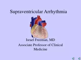
Epidemiology of VA & SCD Classification of Ventricular Arrhythmia by Clinical Presentation • Hemodynamically stable • ♥Asymptomatic • ♥Minimal symptoms, e.g., palpitations • Hemodynamically unstable • ♥ Presyncope • ♥ Syncope • ♥ Sudden cardiac death • ♥ Sudden cardiac arrest
Epidemiology of VA & SCD Classification of Ventricular Arrhythmia by Electrocardiography • Nonsustained ventricular tachycardia (VT) • ♥Monomorphic • ♥Polymorphic • Sustained VT • ♥Monomorphic • ♥Polymorphic • Bundle-branch re-entrant tachycardia • Bidirectional VT • Torsades de pointes • Ventricular flutter • Ventricular fibrillation
Sustained Monomorphic VT 72-year-old woman with CHD
Sustained Polymorphic VTExercise induced in patient with no structural heart disease
Ventricular Flutter Spontaneous conversion to NSR (12-lead ECG)
Wide QRS Irregular Tachycardia:Atrial Fibrillation with antidromic conduction in patient with accessory pathway – Not VT
Epidemiology of VA & SCD Classification of Ventricular Arrhythmia by Disease Entity • Chronic coronary heart disease • Heart failure • Congenital heart disease • Neurological disorders • Structurally normal hearts • Sudden infant death syndrome • Cardiomyopathies • ♥Dilated cardiomyopathy • ♥Hypertrophic cardiomyopathy • ♥Arrhythmogenic right ventricular (RV) • cardiomyopathy
Epidemiology of VA & SCD Incidence of Sudden Cardiac Death Reused with permission from Myerburg RJ, Kessler KM, Castellanos A. Circulation 1992;85:12-10.
Mechanisms and Substrates Mechanisms of Sudden Cardiac Death in 157 Ambulatory Patients • Ventricular fibrillation - 62.4% • Bradyarrhythmias (including advanced AV block and asystole) - 16.5% • Torsades de pointes - 12.7% • Primary VT - 8.3% Bayes de Luna et al. Am Heart J 1989;117:151–9.
Clinical Presentations of Patients with VA & SCD • Asymptomatic individuals with or without electrocardiographic • abnormalities • Persons with symptoms potentially attributable to ventricular • arrhythmias • ♥Palpitations • ♥Dyspnea • ♥Chest pain • ♥Syncope and presyncope • VT that is hemodynamically stable • VT that is not hemodynamically stable • Cardiac arrest • ♥Asystolic (sinus arrest, atrioventricular block) • ♥VT • ♥Ventricular fibrillation (VF) • ♥Pulseless electrical activity
Therapy • Acute • Hemodynamically Stable • Hemodynamically UnStable
Therapies for VA • Antiarrhythmic Drugs • ♥Beta Blockers: Effectively suppress ventricular ectopic beats & arrhythmias; reduce incidence of SCD • ♥Amiodarone: No definite survival benefit; some studies have shown reduction in SCD in patients with LV dysfunction especially when given in conjunction with BB. Has complex drug interactions and many adverse side effects (pulmonary, hepatic, thyroid, cutaneous) • ♥Sotalol: Suppresses ventricular arrhythmias; is more pro-arrhythmic than amiodarone, no survival benefit clearly shown • ♥Conclusions: Antiarrhythmic drugs (except for BB) should not be used as primary therapy of VA and the prevention of SCD
Therapies for VA • Non-antiarrhythmic Drugs • ♥ Electrolytes: magnesium and potassium administration can favorably influence the electrical substrate involved in VA; are especially useful in setting of hypomagnesemia and hypokalemia • ♥ ACE inhibitors, angiotensin receptor blockers and aldosterone blockers can improve the myocardial substrate through reverse remodeling and thus reduce incidence of SCD • ♥ Antithrombotic and antiplatelet agents: may reduce SCD by reducing coronary thrombosis • ♥ Statins: have been shown to reduce life-threatening VA in high-risk patients with electrical instability • ♥ n-3 Fatty acids: have anti-arrhythmic properties, but • conflicting data exist for the prevention of SCD
Hazard ratio Trial Name, Pub Year LVEF, other features N = 196 MADIT-I 0.35 or less, NSVT, EP positive 1996 0.46 AVID N = 1016 Aborted cardiac arrest 1997 0.62 N = 900 0.35 or less, abnormal SAECG and scheduled for CABG CABG-Patch 1.07 1997 N = 191 CASH* Aborted cardiac arrest 2000 0.83 Aborted cardiac arrest or syncope CIDS N = 659 2000 0.82 0.30 or less, prior MI MADIT-II N = 1232 2002 0.35 or less, NICM and PVCs or NSVT 0.69 DEFINITE N = 458 0.35 or less, MI within 6 to 40 days and impaired cardiac autonomic function 2004 0.65 N = 674 DINAMIT 2004 1.08 0.35 or less, LVD due to prior MI and NICM N = 1676 SCD-HeFT 2005 0.77 1.8 1.0 1.2 0.6 0.4 0.8 1.4 1.6 ICDbetter Therapies for VA ICDs: Results from Primary and Secondary Prevention Trials
Therapies for VA • Primary Prevention of SCD (1) • Recommendations in previously published guidelines for prophylactic • ICD therapy based on LVEF are inconsistent: ♥ Different LVEFs were chosen for inclusion in trials ♥ Average EF in such trials was substantially lower than the cutoff value for enrollment ♥ Subgroup analyses in various trials have not been consistent in their implications ♥ No trials contained randomized patients with intermediate LVEF
Therapies for VA • Primary Prevention of SCD (2) • Because of these inconsistencies, the recommendations in this • guideline were constructed to apply to patients with an EF ≤ to a • range of values • The next several slides compare the recommendations of • previously published guidelines with those in this one and the • reasoning behind the writing committee’s decision
Therapies for VA • Primary Prevention of SCD (3) • LV dysfunction due to MI, LVEF ≤ 30%, NYHA class II, III • 2005 ACC/AHA HF: Class I; LOE B • 2005 ESC HF: Class I; LOE A • 2004 ACC/AHA STEMI: Class IIa; LOE B • 2002 ACC/AHA/NASPE PM/ICD: Class IIa; LOE B • 2006 ACC/AHA/ESC VA/SCD: Class I; LOE A • Note: The VA/SCD Guideline has combined all trials that enrolled patients with LV dysfunction due to MI into one recommendation
Therapies for VA • Primary Prevention of SCD (4) • LV dysfunction due to MI, LVEF 30-35%, NYHA class II, III • 2005 ACC/AHA HF: Class IIa; LOE B • 2005 ESC HF: Class I; LOE A • 2004 ACC/AHA STEMI: N/A • 2002 ACC/AHA/NASPE PM/ICD: N/A • 2006 ACC/AHA/ESC VA/SCD: Class I; LOE A • Note: The VA/SCD Guideline has combined all trials that enrolled patients with LV dysfunction due to MI into one recommendation
Therapies for VA • Primary Prevention of SCD (5) • LV dysfunction due to MI, LVEF 30-40%, NSVT, positive EP study • 2005 ACC/AHA HF: N/A • 2005 ESC HF: N/A • 2004 ACC/AHA STEMI: Class I; LOE B • 2002 ACC/AHA/NASPE PM/ICD: Class IIb; LOE B • 2006 ACC/AHA/ESC VA/SCD: Class I; LOE A • Note: The VA/SCD Guideline has combined all trials that enrolled patients with LV dysfunction due to MI into one recommendation
Therapies for VA • Primary Prevention of SCD (6) • LV dysfunction due to MI, LVEF ≤ 30%, NYHA class I • 2005 ACC/AHA HF: Class IIa; LOE B • 2005 ESC HF: N/A • 2004 ACC/AHA STEMI: N/A • 2002 ACC/AHA/NASPE PM/ICD: N/A • 2006 ACC/AHA/ESC VA/SCD: Class IIa; LOE B • Note: The VA/SCD Guideline has expanded the range of LVEF ≤ 30-35% for patients with LVD due to MI and NYHA class I into one recommendation
Therapies for VA • Primary Prevention of SCD (7) • LV dysfunction due to MI, LVEF ≤ 31-35%, NYHA class I • 2005 ACC/AHA HF: N/A • 2005 ESC HF: N/A • 2004 ACC/AHA STEMI: N/A • 2002 ACC/AHA/NASPE PM/ICD: N/A • 2006 ACC/AHA/ESC VA/SCD: Class IIa; LOE B • Note: The VA/SCD Guideline has expanded the range of LVEF ≤ 30-35% for patients with LVD due to MI and NYHA class I into one recommendation
Therapies for VA • Primary Prevention of SCD (8) • Nonischemic cardiomyopathy, LVEF ≤ 30%, NYHA class II, III • 2005 ACC/AHA HF: Class I; LOE B • 2005 ESC HF: Class I; LOE A • 2004 ACC/AHA STEMI: N/A • 2002 ACC/AHA/NASPE PM/ICD:N/A • 2006 ACC/AHA/ESC VA/SCD: Class I; LOE B • Note: The VA/SCD Guideline has combined all trials of nonischemic cardiomyopathy, NYHA class II, III into one recommendation
Therapies for VA • Primary Prevention of SCD (9) • Nonischemic cardiomyopathy, LVEF 30-35%, NYHA class II, III • 2005 ACC/AHA HF: Class IIa; LOE B • 2005 ESC HF: Class I; LOE A • 2004 ACC/AHA STEMI: N/A • 2002 ACC/AHA/NASPE PM/ICD:N/A • 2006 ACC/AHA/ESC VA/SCD: Class I; LOE B • Note: The VA/SCD Guideline has combined all trials of nonischemic cardiomyopathy, NYHA class II, III into one recommendation
Therapies for VA • Primary Prevention of SCD (10) • Nonischemic cardiomyopathy, LVEF ≤ 30%, NYHA class I • 2005 ACC/AHA HF: Class IIb; LOE C • 2005 ESC HF: N/A • 2004 ACC/AHA STEMI: N/A • 2002 ACC/AHA/NASPE PM/ICD:N/A • 2006 ACC/AHA/ESC VA/SCD: Class IIb; LOE B • Note: The VA/SCD Guideline has expanded the range of LVEF to ≤ 30-35% for patients with nonischemic cardiomyopathy and NYHA class I into one recommendation
Therapies for VA • Primary Prevention of SCD (11) • Nonischemic cardiomyopathy, LVEF ≤ 31-35%, NYHA class I • 2005 ACC/AHA HF: N/A • 2005 ESC HF: N/A • 2004 ACC/AHA STEMI: N/A • 2002 ACC/AHA/NASPE PM/ICD: N/A • 2006 ACC/AHA/ESC VA/SCD: Class IIb; LOE B • Note: The VA/SCD Guideline has expanded the range of LVEF to ≤ 30-35% for patients with nonischemic cardiomyopathy and NYHA class I into one recommendation
ICD History The original AID device had two electrodes, one a spring was placed in the Vena Cava, the other a cup designed to conform to the cardiac apex 1980
209 cc 80 cc 113 cc 80 cc 72 cc 54 cc 39.5 cc 36 cc 62 cc 49 cc 39.5 cc 39.5 cc 39 cc 39 cc Medtronic Implantable Defibrillators (1989-2001)
Detection VF Detection ! VF Therapy !! F F F F F F F F F F
Fib Zone 1 2 3 4 5 6 7 8 9 10 11 12 Initiate Therapy Detect Analyse Charge Detection
310 340 300 290 330 320 300 310 Rate Branch Calculation - Example VT Detection: 8 intervals < 350 ms VT Detect 290 300 300 330 340 320 310 310 Median Ventricular Cycle Length = (310 + 310) / 2 = 310 ms
ATP Definition • ATP = Antitachycardia Pacing • ATP = Therapeutic intervention using standard bradycardia pacing algorithms and energy levels in an effort to bring the heart out of a reentrant tachycardia and restore its normal rhythm