Brachial Plexus
Brachial Plexus. Objectives. Describe the brachial plexus Make a list of contributing spinal nerves. Discuss the general arrangement of this plexus. Locate the plexus in the axilla and note important relations to blood vessels. Make a list of the terminal main branches of brachial plexus.


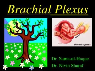
Brachial Plexus
E N D
Presentation Transcript
Objectives • Describe the brachial plexus • Make a list of contributing spinal nerves. • Discuss the general arrangement of this plexus. • Locate the plexus in the axilla and note important relations to blood vessels. • Make a list of the terminal main branches of brachial plexus.
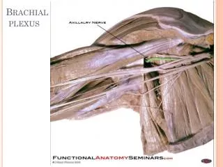
Brachial Plexus is a network of nerves that is present at the root of the neck to enter the upper limb. • Brachial Plexus is present in the posterior triangle of the neck & axilla. • It is formed by the union of the anterior Rami of the • C 5th, 6th, 7th & 8th and the 1st thoracic spinal nerve.
Formation • Roots of C5 & C6 unite to form---- Upper trunk • Root of C7 continuous as the-------- Middle trunk • Roots of C8 & T1 unite to form---- Lower trunk
The Plexus can be divided into5 stages: • Roots: in the posterior∆ • Trunks: in the posterior∆ • Divisions: behind the clavicle (in cervico-axillary canal) • Cords: in the axilla • Branches: in the axilla • The first 2 stages lie in the posterior triangle, while the last 2 sages lie in the axilla. 6
The anterior divisions of the upper and middle trunks unite to form the Lateral cord. • The anterior division of the lower trunk continues as the Medial cord. • All the posterior divisions of three trunks join to form the Posterior cord. 7
CORDS & BRANCHES • Branches • All three cords will give branches, those will supply their respective regions
BRANCHES • (A) From Roots: 1. C5:Nerve torhomboids (dorsal scapular nerve). 2. C5,6 &7:Longthoracic nerve • (B) From Trunks (upper trunk): • Nerve to subclavius • Suprascapularnerve (supplies supraspinatus & infraspinatus)
(C)BRANCHES From Cords Lateral Cord (2LM) .Lateral pectoral n .Lateral root to median n .Musculocutaneous n C5 C6 C7 C8 T1 Medial cord (4MU) .Medial pectoral n. .Medial root to median n. .Medial cutaneous n of arm. .Medial cutaneous n of forearm. .Ulnar n. Posterior Cord (ULTRA) .Upper subscapular n .Lower subscapular n .Thoracodorsal n .Radial n .Axillary n
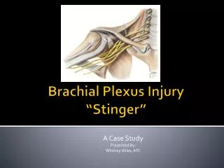
Brachial Plexus Injuries • In Infants: During Difficult Delivery: Shoulder dystocia
Brachial Plexus Injuries • In Adults: • Sports most commonly associated: Football, baseball, basketball, volleyball, wrestling, and gymnastics. • Nerve injuries can result from: Blunt force trauma, poor posture or chronic repetitive stress.
Brachial Plexus Injuries • Patients generally present with pain and/or muscle weakness. • Some patients may experience muscle atrophy.
Erb- Duchenne palsy Damage to the upper trunk: C5, 6 The most commonly involved nerves are the suprascapular nerve, musculocutaneous nerve, and the axillary nerve: paralysis and atrophy of the deltoid, biceps, and brachialis muscles.(supra and infraspinatus) Clinical Appearance: Motor Loss: Arm hangs by side Adducted Shoulder (Deltoid) Medially Rotated Arm (infraspinatus Extended Elbow (brachialis and biceps) Pronated Elbow (biceps) Sensory Loss: Lateral aspect of Upper Limb
klumpke paralysis or Palsy Injury to Inferior part of Plexus (C8, T1) Occurrence: Excessive abduction of arm. Less common than Injury to Superior part of Plexus.
klumpke paralysis or Palsy Clinical Appearance: Motor Loss: Small muscles of Hand:( Interossei, thenar, hypothenar) Flexors of the wrist and fingers: ( Flexor carpi ulnaris, ulnar half of flexor digitorumprofundus) Dilator pupillae, Levatorpalpebraesuperioris (T1): Hornerssyndrome Sensory Loss: Medial aspect of Upper Limb
Cervical Rib Involves Inferior part of Plexus
What is Waiter’s tip or Porter’s tip position?