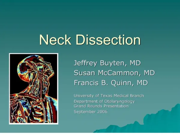
Neck Dissection
Objectives. Natural history of metastatic neck diseaseTumour biologyOccult neck diseaseExtracapsular spreadClinical stagingCT/MRI/USS/PETOpen biopsy vs. sentinal nodeDifficult neck

Neck Dissection
E N D
Presentation Transcript
1. Neck Dissection Stephen Ball
2. Objectives Natural history of metastatic neck disease
Tumour biology
Occult neck disease
Extracapsular spread
Clinical staging
CT/MRI/USS/PET
Open biopsy vs. sentinal node
Difficult neck & contraindications
Neck dissections
3. Introduction Over 500 lymph nodes in the body
200 of these in the head & neck
Normally 3mm � 3cm, most <1cm
Many H&N tumours will undergo nodal spread
Presence, absence, level & size of metastatic neck disease of significant prognostic determinant
Literature confusing
Retrospective analyses
Non-randomised
Selection bias
Survival/locoregional control endpoint
4. Natural history of Neck disease Key factors
Tumour site
Tumour size
Tumour thickness
<5mm 16% LN +ve, >5mm 64% LN +ve
Previous treatment
Tumour recurrence
Tumour histology
Tumour immunology
5. Primary tumour site predictable based on distribution of cervical metastasis*
Memorial Sloan-Kettering levels*
7. Organ specific drainage
9. Tumour biology Are lymph nodes favourable site for tumour growth
Limitless replication vs. tumourlysis
Cancer cells ? Lymphatic system via endothelial gaps
Passive transport in lymph
2-4g tumour 4x10⁶ cells/g/day*
Anti-tumour/filtering function poorly understood
10. SCC growth patterns within cervical LN*
Subcapsular deposits growth within node ++ ? extranodal spread via capsular disruption
Early extranodal spread from intranodal growth
Malignant embolus ? subcapsular sinus + capsular lymphatics ? intra + extra nodal disease
Only capsular embolus no intranodal disease ? early extranodal spread
11. Stages of lymphatic metastasis
Premetastatic invasion of tumour epithelial basal lamina
Penetration of lamina
Translocation of tumour cells through a lymphatic
Exit from node
Venous drainage
Lymphatic drainage
Direct spread
12. Molecular detection of metastases Histologically normal tissue ? absence of tumour
Molecular assays > 500x sensitive*
Micro-array
QRT-PCR
Oligonucleotide mismatch assay
Mitochondrial DNA mutations
13. Occult nodal disease N0 N+ Neck ~25%
Pathologically +ve nodes in 30% elective neck dissection*
Occult neck disease can = subsequent clinical disease
Subclinical spread ? early cancer
Can only detect occult disease on removal
Patients with micrometastasis 3x more curable than those with macroscopic disease*
Literature currently does not justify discovering occult nodal disease on a routine basis
N0 neck + risk occult mets from 10 site >20% consider SND
50% risk antr tongue, oropharynx & hypopharynx
14. Extracapsular spread General consensus extracapsular spread = poor prognosis*
Soft tissue invasion ?success by >80%
Occult nodal disease & extracapsular spread poor prognosis
No properly controlled prospective study comparing survival to extracapsular spread
High risk patients (+ve resection margin, extranodal spread, perineural involvement) improved overall survival & locoregional control when treated with post op combined chemoradiation*
? tumour burden vs. ? tumour aggressiveness
? Depressed host-immune response?
15. Clinical Staging UICC/AJC classification for regional cervical lymphadenopathy*
Applies to all H&N tumours except nasopharynx & thyroid
Criticisms
Most important prognostic factors thought to be no. of nodes + extracapsular spread � neither can be measured clinically
Clinical stage emphasises laterality
Bilateral nodes ? worse prognosis eg. N1 supraglottis
No independent classification of massive bilat nodes, often fixed & universally fatal.
16. CT More accurate than clinical examination
647 neck dissections
Sensitivity 84%
Specificity 83%
Clinical examination
Sensitivity 74%
Specificity 81
Especially useful in difficult necks: restaging, retropharynx
As cancer invades the node
Enlarges
Necrotic centre
Peripheral inflammation = rim enhancement
CT nodes >1cm ~80% accuracy
low-level II & high level III >1.5cm
Difficulties: low-volume neck disease + residual/recurrent disease following surgery & irradiation
17. MRI Similar accuracy to CT
Size criteria similar
Maybe better in evaluating N0 neck
Window settings ~helpful in identifying malignant nodes
Superparamagnetic iron oxide (SPIO) used as lymphangiographic agents
Taken up up by RES in normal & inflammed nodes ? signal drop off
No signal change in metastaic nodes
18. Ultrasound Detect presence of cervical nodes
Ability to differentiate malignant vs. benign limited
Sensitivity ~75-95%
Specificity ~63-91%
Can be combined with FNA
19. Radionuclide & PET Radionuclides e.g. Gallium-67, Technetium-99 dimercaptosuccinic acid (DMSA)
Low sensitivity/specificity
Inability to detect low volume disease
PET � assess metabolic activity of nodes using 18 fluorodeoxyglucose (FDG)/
Still poor sensitivity/specificity for low volume disease
CT/PET useful for:
occult 10
Residual/recurrent disease following surgery & radiotherapy*
20. Sentinal node Non-H&N melanoma + breast carcinoma
Not routinely used:
Exact nature of H&N lymphatic drainage unclear
Skip metastasis do occur
Collatral channels often present
Necessitates operating in oncologically significant area
Facial nerve risk in parotid nodes
Learning curve & operator dependent
Role limited to T1N0 oral cavity & oropharynx
RCT by EORTC pending
21. Open biopsy Generally avoided
Equivocal cases/lymphoma/anaplastic carcinoma/FNAC not available
No evidence in literature open Bx alters prognosis
Provided correct treatment instigated within six weeks*.
Incision should be planned to facilitate scar removal by subsequent standard neck dissection incision
22. Difficult Neck Difficult to access
Short stocky neck
Recurrent disease
Retropharyngeal nodes
Extensive disease around vital structures
Brachial plexus, prevertebral muscles, carotid
Pre-op planning e.g. risk of hemiplegia by assessing collateral supply from circle of Willis.
23. Contraindications Absolute vs. relative
Primary tumour untreatable
Medically unfit for anaesthetic
Inoperable neck disease
Fixation to skull base/brachial plexus
Distant metastasis
Radical radiotherapy/+- adjuvant chemotherapy/symptom palliation
24. Recurrence & salvage surgery Poor prognosis
50% chance salvaging recurrent cancer in untreated neck
25% in electively irradiated neck
5% previously dissected neck*
25. Neck Dissection 1906 Crile described classic radical neck dissection
Popularised by Hayes Martin
Comprehensive
removal of all five lateral lymph node levels
Selective
26. Incision
27. Radical neck dissection Removal of LN containing levels I-V
All 3 non-lymphatic structures
SAN
SCM
IJV
28. Extended radical Radical neck dissection plus :
One or more LN groups
Retropharyngeal LN
Parpharyngeal LN
Parotid LN
Level VI/VII LN
Non-lymphatic structures
Mandible
Parotid
Mastoid tip
Prevertebral fascia & musculature
Digastric
Hypoglossal n.
External carotid
Skin
Or both
29. Modified radical neck dissection Removal of all level I-V LN with preservation =1 non-lymphatic structure
Type 1 � SAN
Type 2 � SAN & IJV
Type 3 � SAN & IJV & SCM (functional)
30. Selective neck dissection