Basic Echocardiography
Basic Echocardiography. Wendy Blount, DVM Nacogdoches TX. Echo Technique - Anatomy. Tricuspid valve Septal leaflet Parietal leaflet Pulmonic Valve Right cusp Left cusp Intermediate cusp. Mitral valve Leaflets are less distinct Aortic Valve Right cusp Left cusp Septal cusp.

Basic Echocardiography
E N D
Presentation Transcript
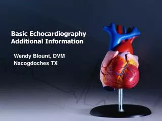
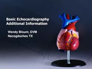
Basic Echocardiography Wendy Blount, DVM Nacogdoches TX
Echo Technique - Anatomy Tricuspid valve • Septal leaflet • Parietal leaflet Pulmonic Valve • Right cusp • Left cusp • Intermediate cusp Mitral valve • Leaflets are less distinct Aortic Valve • Right cusp • Left cusp • Septal cusp
Echo Technique - Anatomy RV • Conus arteriosus • 3 papillary muscles LV • 2 papillary muscles
Echocardiography Equipment • Transducer – small footprint • Fan-shaped beam or sector • High frequency for small animals • Low frequency for large animals • Machines range from 2.5-10 Mhz • 5-7 MHz will work fine for most dogs and cats for echo
Echocardiography Equipment • Double window with simultaneous B and M modes (video) • Can do measurements on B-mode or M-mode • Need a cursor which can measure mm, or cm marks on the images • Ability to capture images is important
Echocardiography Preparation • Thin coated animals – alcohol, part the hairs, gel • Thick coated animals – shave the window – at the sternum, just behind the elbow • Sedation only if needed • Acepromazine – 0.025 mg/lb (max 1 mg) • Buprenex – 0.01-0.02 mg/kg • Mix together and give IV (handout)
Echocardiography Positioning for 8 standard views • Right lateral recumbency • Cardiac table is nice but not necessary • Sonographer needs a stool or chair • Placement of probe: • Feel the apical beat, and put your probe there (probe marker cranial) • Imagine the longitudinal axis of the heart, probe at 90o (short axis views) • Adjust 1 intercostal space Cr or Cd PRN • Rarely move the probe head – just fan and twist (video)
1. Short Axis – Left Ventricle • Fan from base to apex, until you have just passed the mitral valve, and the LV papillary muscles appear (mushroom view) • Rotate until PM are the same size • If you are getting a rib shadow, try one intercostal space cranial or caudal • Fan cranial and caudal to center the heart on the screen
1. Short Axis – Left Ventricle Abbreviations - Structures • P – pericardium • RV – right ventricle • IVS – intraventricular septum • LV – left ventricle • PPM – posterior papillary muscle • APM – anterior papillary muscle
1. Short Axis – Left Ventricle Measurements • IVSTd- IntraVentricular Septum Diastole • LVIDd - LV Inner Diameter Diastole • LVPWd – LV Posterior Wall Diastole • IVSTs- IntraVentricular Septum Systole • LVIDs - LV Inner Diameter Systole • LVPWs – LV Posterior Wall Systole
1. Short Axis – Left Ventricle Measurements • IVSTd =IVSd =VSd • LVIDd=LVd =LVLd • LVPWd=LVFWd=LVWd • IVSTs =IVSs =VSs • LVIDs=LVs =LVLs • LVPWs=LVFWs=LVWs
1. Short Axis – Left Ventricle Measurements - Calculated • FS – fractional shortening (LVIDd – LVIDs) LVIDd • Assumes perpendicular to myocardium • Assumes contractility is uniform in the LV • Extremes in preload and afterload can affect FS, as well as myocardial function
1. Short Axis – Left Ventricle Measurements - Calculated • FS – fractional shortening • AKA shortening fraction (SF) • >30% in the dog • >40% in the cat • >45% if MR is compensated
1. Short Axis – Left Ventricle Measurements - Tips • Make sure you don’t include PM in the LVPW measurement • If you do, your LVPW will be artifactually thicker • Clue – check for this if LVPW is much thicker than IVS • Make sure you are not too far apical • If you are, your LVID will be artifactually small • And LVPW will be artifactually thick
1. Short Axis – Left Ventricle Measurements - Tips • Measure three times • Take the average • Throw out any outliers • Several sets of normals published • 1-2mm outside normal may not always be significant
2. Short Axis – Apex Structures • Pericardium • May or may not see RV • LV apical lumen No measurements here
3. Short Axis – Chordae Tendinae Structures • Pericardium • RV • LV • CH - Chordae Tendinae (posterior & anterior) No measurements here
4. Short Axis – Mitral Valve Structures • Pericardium • RV • RV Papillary Muscles • LV • MV - Mitral Valve (Posterior & Anterior)
4. Short Axis – Mitral Valve Measurement • EPSS – E-Point to Septal Separation • Can denote decreased LV systolic function • Less than 6 mm in large dogs • Less than 3-5 mm in small dogs and cats
5. Short Axis – Aortic Valve Structures • RVOT – Right Ventricular Outflow Tract • TV – Tricuspid Valve • PV – Pulmonic Valve • Ao – Aortic Valve • LA – Left Atrium
5. Short Axis – Aortic Valve Measurements • Ao – at largest dimension (systole) • LA – at largest dimension (diastole) • LA:Ao – • 0.8 to 1.3 in dogs • 0.8 to 1.4 in cats
6. Short Axis – Pulmonary Artery Structures • RA – Right Atrium • Ao – Aorta (ascending) • PA– Pulmonary Artery • LPA – left pulmonary artery • RPA – right pulmonary artery • CaVC – Caudal Vena Cava
7. Long Axis – 4 Chamber Technique • Get short axis “mushroom” view • Rotate 90 degrees counterclockwise
7. Long Axis – 4 Chamber Structures • RV – Right Ventricle • RA – Right Atrium – difficult to view completely • TV – Tricuspid Valve • LV – Left Ventricle • LA – Left Atrium • MV – Mitral Valve, PM – papillary muscle
7. Long Axis – 4 Chamber Video
8. Long Axis – LVOT Technique • Find 4 Chamber view • Angle the “dot” toward the shoulders • Elevate the cord end of the probe
8. Long Axis – LVOT Structures • RV, TV, RA • LV, PM, MV • Very edge of the LA • LVOT – AV (LC, SC), ascending Ao • RPA – Right Pulmonary Artery
8. Long Axis – LVOT Video Normal Dog Video
Dog RV Measurement Values • RVWd – less than LVWd • RVIDd – 1/3 or less of LVIDd (handout)
Cat Echo Normal Values • IVSTd – 3-6 mm • LVIDd – 10-21 mm • LVPWd – 3-6 mm • IVSTs - 4-9 • LVIDs – 4-11 mm • LVPWs – 4-10 mm • Aos – 6-12 mm • LAd – 7-15 mm • FS - >40% • EPSS - 0-3 mm • EF ->70% • LA:Ao – 0.8-1.4 • RVIDd - 3-7 mm • RVWd - <3 mm (form)
Ferret Echo Normal Values (Mean) • LVIDD – 11.0 mm • LVIDS - 6.4 mm • LVPW - 3.3 mm • FS - 42% • EPSS - 0