Download

1 / 54
540 likes | 763 Views
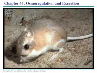
Excretion and Osmoregulation. EXCRETION IS THE S**T!. By: Julia Messineo, Haley Giangreco & Nora Shafer. Introduction from Brain Pop!. http://www.brainpop.com/health/bodysystems/urinarysystem /. 44.1 – Osmoregulation Balances the Uptake and Loss of Water and Solutes. A Balancing Act:

E N D
Excretion and Osmoregulation EXCRETION IS THE S**T! By: Julia Messineo, Haley Giangreco & Nora Shafer
Introduction from Brain Pop! http://www.brainpop.com/health/bodysystems/urinarysystem/
44.1 – Osmoregulation Balances the Uptake and Loss of Water and Solutes • A Balancing Act: • The physiological system of animals (cells/tissues) (organs/organ systems) operate in a fluid environment. • Proper functions of these systems depend on relative concentrations of water and solutes • Functionality is challenged by animal's external environment • Ex./ Freshwater animals live in an environment that threatens to flood and dilute their body fluids, these animals show adaptions that reduce water uptake, conserve solutes, and absorb salts from their surroundings. Success in environments depend on these adaptations. • Animals maintain homeostatic processes by: • Osmoregulation: how animals regulate solute concentrations and balance the gain and loss of water • Excretion: how animals get rid of nitrogen-containing waste products of metabolism
44.1 – Osmoregulation Balances the Uptake and Loss of Water and Solutes • Animals abilities to regulate the chemical composition of its body fluids depends on balancing the uptake and loss of water and solutes. • This osmoregulation is based largely on controlled movements of solutes between internal fluids and the external environment. • This process also regulates the movement of water, which follows solutes by osmosis. • Animals remove metabolic waste products before they reach harmful levels.
Osmosis • ALL ANIMALS, regardless of type or waste products, face the same central problem of osmoregulation • Rates of water loss and uptake must balance • Animal cells swell and burst if water uptake is continuous • Animal cells shrivel and die if water loss is continuous
Osmotic Challenges • There are two basic problems of balancing water gain with water loss: • One (only available to marine animals) is to be isoosmotic to surroundings • Two solutions separated by a selectively permeable membrane have the same permeable membrane • Osmoconformer: animal that does not actively adjust its internal osmolarity • Because internal osmoregularity is the same as its environment, there is no tendency to gain or lose water • Live in water with stable composition and have a very constant internal osmolarity • Osmoregulator: (in contrast) an animal that must control its internal osmolarity because its body fluids are not isoosmotic with the outside environment • Must discharge excess water if it lives in a hypoosmotic environment or take in water to offset the loss • More dilute solution • Allows animals to maintain internal osmolarities different from that of their environments
Osmotic Challenges • Whenever animals maintain an osmoregularity difference between the body and the external environment there is an energy cost • Animals must expand energy to maintain the osmotic gradients that cause water to move in and out • Active transport is used to manipulate solute concentrations in their body fluids • Most animals cannot tolerate substantial changes in external osmolarity and are said to be stenohaline • Referring to salt • Uryhaline: can survive large fluctuations in external osmolarity
Marine Animals • Even an animal that conforms to the osmolarity of its surroundings regulates its internal composition of solutes • Osmoregulation in a saltwater fish: • Gain of water and salt ions from food and by drinking seawater • Excretion of salt ions from gills • Osmotic water loss through gills and other parts of body surface • Excretion of salt ions and small amounts of water in scanty urine from kidneys
Freshwater Animals • Osmoregulation in a freshwater fish: • Uptake of water and some ions in food • Uptake of salt ions by gills • Osmotic water gain through gills and other parts of body surface • Excretion of large amounts of water in dilute urine from kidneys
Animals That Live in Temporary Waters • Some animals can lose almost all of their body water and live in a dormant state when their habitats dry up (anhydrobiosis) • These animals can survive on 2% water and live in an inactive state for a decade or more • Just add water and within minutes the rehydrated tardigrades are moving about and feeding • These dehydrated individuals contain large amounts of sugars and seem to protect the cells by replacing the water that is normally associated with membranes and proteins
Land Animals • Mammals that evolved in dry environments can withstand up to 24% loss of water in the body • The waxy cuticle helps plants prevent water loss • Nocturnal animals lose less water by taking advantage of the lower temperature and higher relative humidity of night air • Despite these adaptations most terrestrial animals lose considerable water from moist surfaces in their gas exchanges, organs, urine, feces, and across their skin • Land animals balance this loss by eating and drinking moist foods and by using metabolic water (water produced during cellular respiration)
Transport Epithelia • The ultimate function of osmoregulation is to maintain the composition of cellular cytoplasm • Most animals do this indirectly by managing the composition of an internal body fluid that bathes the cells • In insects and other animals with an open circulatory system this fluid is the hemolymph • In vertebrates and other animals with a closed circulatory system the cells are bathed in an interstitial fluid that is controlled indirectly through the composition of blood • This fluid composition is maintained by specialized structures coming from cells that regulate solute movement to complex organs such as the vertebrate kidney
Transport Epithelia • Transport epithelium: layers of specialized epithelium cells and regulate solute movements are essential movements and components of osmotic regulation and metabolic waste disposal • Some face the outside environment directly while some face the inside of the body • This arrangement ensures that any solutes moving between animal and environments must pass through a selectively permeable membrane • Ex./ salt glands in marine birds (albatross) • The molecular structure of the plasma membrane determines the kinds and directions of solutes that move across a particular type of transport epithelium • Transport epithelium in excretory organs often have the dual functions of maintaining water balance and disposing of metabolic wastes
44.2 – An Animal’s Nitrogenous Wastes Reflect Its Phylogeny and Habitat • Animals waste products have a large impact on its water balance • Most important waste products are the nitrogenous breakdown of products of proteins and nucleic acids • When these macromolecules are broken apart for energy or converted to carbohydrates and fats enzymes remove nitrogen in the form of ammonia • There are multiple ways ammonia is excreted, mainly directly. But mangy species convert ammonia to other, less toxic, compounds that further require every, ATP, to produce.
Forms of Nitrogenous Waste • Different animals excrete nitrogenous waste in differing forms, varying in toxicity and energy costs • Ammonia • Animals that excrete this need lots of access to water, making it most common in aquatic species • Ammonia molecules can easily pass through membranes and are readily lost by diffusion to the surrounding water • Urea: substance produced in the vertebrate liver by a metabolic cycle that combines ammonia with carbon dioxide • Works well in aquatic species but is much less suitable for land animals • Can only be excreted in very dilute solutions, most land animals do not have the access to water that would achieve that • ADVANTAGE: low toxicity, permits transport by animals safely, animals require much less water because not as much is needed to dilute the solution • DISADVANTAGE: animals must expend energy to produce urea from ammonia
Forms of Nitrogenous Waste • Uric Acid: major nitrogenous waste of insects, land snails, reptiles, and birds • Largely insoluble in water • Can be excreted as a semi solid paste with very little water loss • COST: take a massive amount of energy to produce (more than Urea and more ATP synthesis than ammonia)
The Influence of Evolution and Environment on Nitrogenous Wastes • The kinds of nitrogenous wastes excreted depend on the animals evolutionary history and habitat (especially the availability of water) • The model of reproduction is important in determining whether uric acid or urea evolves • Ex./ shelled eggs and uric acid • Evolution determines the limits of physiological responses for a species but during their lives individual organisms make physiological adjustments within these evolutionary constraints • The amount of nitrogenous waste produced depends on what kind of food the animal eats-the energy budget • Energy used at high rates means the eating of more food and more nitrogenous wastes being produced • Predators excrete more nitrogen than animals that rely mainly on lipids or carbohydrates as energy sources
44.3 – Diverse Excretory Systems are Variations On a Tubular Theme Regulation of solute movement between internal fluids and external environments determine how the problems regarding water balance are solved Excretory systems, which are the central to homeostasis, dispose metabolic wastes and control body fluid composition by adjusting rate of solute loss
Excretory Processes • All excretory systems produce fluid waste urine in a process involving several steps • 1. Filtration – The excretory tubule collects a filtrate from the blood • Water and solutes are forced by blood pressure across the selectively permeable membranes of a cluster of capillaries and into the excretory tubule • 2. Reabsorption – the transport epithelium reclaims valuable substances from the filtrate and returns them to the body fluids • 3. Secretion – other substances, such as toxins and excess ions, are extracted from body fluids and added to the contents of the excretory tubule • Excretion – the filtrate leaves the system and body • *Key functions of excretory system: produce a filtrate by pressure-filtering body fluids and modifying filtrate’s contents
Survey of Excretory Systems • Systems which perform general excretory functions vary among different animal groups • Built on a complex network of tubules • Provides a large surface area for the exchange of water and solutes (nitrogenous wastes)
Protonephridia: Flame-Bulb Systems • This is the type of excretory system found in flatworms • Protonephridium – network of dead-end tubules lacking internal openings • Tubules branch throughout the body, and the smallest branches are capped by the flame bulb (cellular unit) • Flame bulb has a tuft of cilia projecting in tubule • Beating of cilia draws water/solutes from interstitial fluid through flame bulb (filtration) into tubule system • The moves urine outward through tubule until emptied completely into external environment called nephridiopores • Excreted urine is dilute in freshwater flatworms, and helps balance osmotic uptake of water from environment • Flame-bulb systems of freshwater flatworms function solely in osmoregulation • Difference in function shows that structure common to a group adapt in diverse ways by evolution in difference environments
Metanephridia • Has internal openings that collect body fluids • Found in most annelids • Ex/ Earthworms • Each segment of worm has a pair of metanephridia – immersed in coelomic fluid and enveloped by capillary network • Internal network surrounded by ciliated funnel called the nephrostome • Fluid enters nephrostome through collecting tubule, including storage bladder that opens through the nephridiopores • Excretory/ osmoregulatory functions: • As urine progresses though tubule, transport epithelium bordering lumen reabsorbs majority of solutes and returns them to blood in capillaries • Nitrogenous wastes remain in tubule then excrete to outside • Metanephridia of earthworms balance water influx through the production of dilute urine
Malpighian Tubules • Found in insects and other terrestrial anthropods • Remove nitrogenous wastes and function in osmoregulation: • Open in digestive tract and dead-end that are immersed in the circulatory fluid (hemolymph) • Solutes are secreted through transport epithelium lining • Water follows the solutes into tubule through osmosis, fluid passes to rectum (solutes pumped back into hemolymph), and water follows solutes and nitrogenous wastes (soluble uric acid) are eliminated with feces • Insect excretory system is one of the major adaptations contributing to the success of land animals due to its ability to conserve water in an effective manner
Vertebrate Kidneys Usually function in both excretion and osmoregulation Built of tubules Because osmocomforting hagfishes are invertebrates yet have kidneys with segmented excretory tubules, it is suggested that vertebrate ancestors had segmented excretory structures Compact, non-segmented organs consisting of multiple tubules Dense network of capillaries associated with tubules, ducts, and other structures that carry urine out of tubules and kidneys, are integral parts of vertebrate excretory system
44.4 – Nephrons and Associated Blood Vessels are the Functional Units of the Mammalian Kidney Excretory system of mammals focuses on kidneys Kidneys are the principal site of water balance and salt regulation Mammals have a pair of kidneys Each kidney is bean-shaped, 10 cm long in humans, supplied blood by the renal artery and drained by a renal vein Blood flow is voluminous Kidneys account for less than 1% of body weight yet receive roughly 20% of resting cardiac output Urine exits each kidney through a duct (ureter) and both ureters drain into urinary bladder During urination, urine is expelled from urinary bladder through a tube known as the urethra, which is then emptied to outside near vagina (females) or penis (males) Urination is regulated by sphincter muscles near junction of urethra and bladder (nervous system)
Structure and Function of the Nephron and Associated Structures • Mammalian kidney has two distinct regions: • Outer renal cortex • Inner renal medulla • Microscopic excretory tubules and associated blood vessels pack both regions of kidney • Nephron (functional unit of vertebral kidney) contains a single long tubule and ball of capillaries (glomerulus) • Blind end of tubule forms cup-shaped swelling surrounding glomerulus (Bowman’s capsule) • Human kidney contains roughly one million nephrons (tubule length of 80km)
Filtration of the Blood • Occurs as blood pressure forces fluid from blood in glomerulus into lumen of Bowman’s capsule • Porous capillaries and specialized capsule cells (podocytes) are permeable to water and small solutes but not to blood cells or large molecules • Ex/ plasma proteins • Filtration of small molecules is nonselective • Filtrate in Bowman’s capsule contains: salts, glucose, amino acids, vitamins, nitrogenous wastes (urea), and other small molecules
Pathway of the Filtrate • Starting at Bowman’s capsule, filtrate passes through three regions of nephron: • Proximal tubule • Loop of Henle – hairpin turn with descending and ascending limb • Distal tubule – empties into a collecting duct, receiving processed filtrate from numerous nephrons • Filtrate flows from many collecting ducts of kidney into the renal pelvis which is drained by the ureter • In human kidney, 80% of nephrons (cortical) have reduced loops of Henle and are confined to renal cortex • Remaining 20% consists of juxtamedullary nephrons which have well-developed loops extending into renal medulla • Only mammals and birds have juxtamedullary nephrons • Nephrons of other vertebrates lack loops of Henle • Juxtamedullary nephrons enable mammals to produce urine that is hyperosmotic to body fluids • Extremely important for water conservation • Nephrons and collecting duct are lined by transport epithelium process filtrate to form urine • Task: reabsorption of solutes and water
Blood Vessels Associated With the Nephrons • Each nephron is supplied with blood by an afferent arteriole • A branch of the renal artery that subdivides into the capillaries of the glomerulus • Capillaries converge as leaving glomerulus, forming efferent arteriole • Vessel subdivides again, forming peritubular capillaries, which surround the proximal and distal tubules • More capillaries extend downward and form vasa recta, AKA the capillaries that serve loop of Henle • Vasa recta forms loops with descending and ascending vessels transferring blood in opposite directions • Excretory tubules and surrounding capillaries are closely associated • Do not exchange materials directly • Tubules and capillaries immerse in interstitial fluid – numerous substances diffuse between plasma within capillaries and filtrate within nephron tubule • Exchange facilitated by relative direction of blood flow and filtrate flow in nephrons
From Blood Filtrate to Urine: A Closer Look Filtrate becomes urine as it flows through mammalian nephron and collecting duct
1. Proximal Tubule • Secretion and reabsorption alter volume and concentration of filtrate • Ex/ cells of transport epithelium maintain relativity constant pH in body fluids by controlled secretion of H+ • Cells synthesize and secrete ammonia neutralizes acid from becoming too acidic • The more acidic the filtrate, the more the cells produce ammonia from source • Reabsorb 90% of the buffer bicarbonate • Drugs/poisons are processed in liver and pass from peritubular capillaries into interstitial fluid, and are then secreted across epithelium of proximal tubule into nephron’s lumen • Valuable nutrients including glucose, amino acids and potassium are actively or passively transported from filtrate to interstitial fluid, then moved to peritubular capillaries • Important function of proximal tubule: reabsorption of NaCl and water from huge initial filtrate volume
2. Descending Limb of the Loop of Henle • Reabsorption of water continues as filtrate moves into descending limb • Transport epithelium is freely permeable to salt/small solutes • Interstitial fluid bathing tubule must be hyperosmotic to filtrate • Osmoregularity of interstitial fluid becomes greater from outer cortex to inner medulla • Filtrate moving downward from cortex to medulla within descending limb of loop of Henle loses water to interstitial fluid of greater osmolarity increase of solute concentration in filtrate
3. Ascending Limb of the Loop of Henle • Filtrate reaches tip of lop deep in renal medulla in case of juxtamedullary nephrons • Moves back to cortex with ascending limb • Transport epithelium is permeable to salt but not water* • Two specialized regions: • Thin segment near loop tip – as filtrate ascends, NaCl (concentrated from descending limb) diffuses out of permeable tubule into interstitial fluid • Increases osmolarity of interstitial fluid in medulla • Thick segment adjacent to distal tubule – as salt exits from filtrate, it continues here, but epithelium actively transports NaCl into interstitial fluid • Losing salt without giving up water filtrate dilutes as it moves up cortex in ascending limb
4. Distal Tubule Plays key role in regulating Potassium and NaCl concentrations of body fluids by varying amount of potassium secreted into filtrate and amount of NaCl reabsorbed from filtrate Contributes to pH regulation by the controlled secretion of H+ and reabsorption of bicarbonate
5. Collecting Duct • Carries filtrate through medulla to renal pelvis • Active reabsorption of NaCl allows transport epithelium of collecting duct to play role in determining how much salt is excreted in urine • Epithelium is permeable to water • Not permeable to salt, or in the renal cortex, permeable to urea • Collecting duct transverses gradient of osmolarity in kidney, and filtrate becomes increasingly concentrated as it loses water from osmosis to the hyperosmotic interstitial fluid • Duct in inner medulla becomes permeable to urea • Due to high urea concentration in filtrate, urea diffuses out of duct and into interstitial fluid • Urea, with NaCl, contributes to high osmolarity of interstitial fluid in medulla • High osmolarity enables mammalian kidney to conserve water by excreting urine hyperosmotic to general body fluids
44.5 – The Mammalian Kidney’s Ability to Conserve Water is a Key Terrestrial Adaptation The mammalian kidney can produce urine much more concentrated than body fluids, allowing the conservation of more water.
Solute Gradients and Water Conservation • cooperative action and precise arrangement of the loops of Henle and the collecting ducts are essential for the osmotic gradients that concentrate the urine. • nephrons can be seen as little energy-consuming machines whose function is to produce a region of high osmolarity in the kidney it then extracts water from the urine in the collecting duct • Primary solutes in this osmolarity gradient are NaCl and urea. • Juxtamedullary nephrons are key to the awareness of the physiology of the mammalian kidney as a water-conserving organ • The countercurrent multiplier system of the loop of Henle • Although the two limbs of the loop are close enough to exchange substances through the interstitial fluid • This maintains a high salt concentration in the interior of the kidney, enabling the kidney to form concentrated urine • The countercurrent system of the vasa recta has descending and ascending vessels carrying blood in opposite directions through the kidney’s osmolarity gradient
Solute Gradients and Water Conservation • Can supply the kidney with nutrients and other important substances without interfering with the osmolarity gradient necessary to excrete a hyperosmotic urine • The countercurrent-like characteristics of the loop of Henle and the vasa recta maintain the steep osmotic gradient between the medulla and the cortex • This active transport requires the kidney to have one of the highest metabolic rates of the organs • The collecting duct is permeable to water but not salt • Water leaves by osmosis into the hyperosmotic interstitial fluids, but salts cannot diffuse • Concentration of salt, urea, and other solutes in the filtrate. • Urea diffuses out of the lower portion of the collecting duct, adding to the high osmolarity of the inner medulla • High osmolarity allows the solutes in the urine to be secreted with minimal • water loss • The juxtamedullary nephron is a key adaptation to terrestrial life, enabling mammals to get rid of salts and nitrogenous wastes without squandering water
Regulation of Kidney Function • Mammalian kidney can adjust both the volume and osmolarity of urine depending on the animal’s water and salt balance and the rate of urea production. • With high salt levels and low water amount, a mammal can excrete urea and salt with minimal water loss in small volumes of hyperosmotic urine. • With low salt level and high fluid intake, the kidney can get rid of excess water with little salt loss by producing large volumes of hypoosmotic urine • The adaptability of osmoregulatory function is controlled by nervous and hormonal controls.
Antidiuretic Hormone (ADH) • Important in regulating water balance • ADH is produced is released from the pituitary gland when blood osmolarity rises above a set point and it reaches the kidney. • Osmoreceptor cells in the hypothalamus monitor the osmolarity of the blood. • Causes the distal tubules and collecting ducts to become more permeable to water • Increased water reabsorption. • Urine volume is reduced, preventing increase of blood osmolarity above the set point again. • Osmolarity of the blood decreases the osmoreceptor cells activity in the hypothalamus by negative feedback , and less ADH is secreted.
Antidiuretic Hormone (ADH) • On the contrary, when a large intake of water has reduced blood osmolarity below the set point, a smaller amount of ADH is released. • The permeability of the distal tubules and collecting ducts is lowered, therefore water reabsorption reduces causing an increased excretion of dilute urine. • Alcohol can inhibit the release of ADH and disturb water balance, leading to excessive urinary water loss and dehydration (this contributes the symptoms of a hangover). • Blood osmolarity, ADH release, and water reabsorption in the kidney are all linked in a feedback loop that help in conserving homeostasis.
Juxtaglomerular Apparatus (JGA) • Regulatory mechanism that is a special tissue located near the afferent arteriole that supplies blood to the glomerulus. • When blood pressure or blood volume drops, an enzyme called renin starts chemical reactions that change angiotensinogen (a plasma protein) to angiotensin II (a peptide). • Angiotensin II increases blood pressure and blood volume • Constricts arterioles it increases blood pressure in decreasing blood flow to capillaries, esp. in the kidney. • Stimulates the proximal tubules to reabsorb more Sodium Chloride and water. • Stimulates the adrenal glands to release the hormone aldosterone • This causes distal tubules to reabsorb Na+ and water
Juxtaglomerular Apparatus (JGA) • The renin-angiotensin-aldosterone system (RAAS) is part of a complex feedback circuit that functions in homeostasis • ADH and RAAS increase water reabsorption, they counter different osmoregulatory problems • The release of ADH is a response to an increase in the osmolarity of the blood • When the body is dehydrated from excessive loss or not enough intake of water • The RAAS will detect the fall in blood volume and pressure increasing water and Na+ reabsorption. • ADH and the RAAS are partners in homeostasis. • ADH would lower Na+ concentration in blood by causing water reabsorption in the kidney, but RAAS helps maintain equilibrium by causing Na+ reabsorption.
Atrial Natriuretic Factor (ANF) • Hormone that opposes the RAAS. • The walls of the atria release ANF due to an increase in blood volume and pressure • ANF inhibits the excretion of renin from the JGA, inhibits NaCl reabsorption by the collecting ducts, and reduces aldosterone release • This lowers blood pressure and volume • ADH, the RAAS, and ANF together provide a system of checks and balances that regulates the kidney’s ability to control the osmolarity, salt concentration, volume, and pressure of blood
Desmodus Rotundas • The South American vampire bat, exemplifies the flexibility of the mammalian kidney to adapt to different osmoregulatory and excretory problems • These bats fly long distances find suitable prey so they must consume so much blood that they would be too heavy to fly • The bat’s kidneys excrete lots of dilute urine as it feeds to lose a majority of the water absorbed from a blood meal, and they can fly away • Back in the roost (nest), the bat faces another regulatory problem where most of its food is protein causing lots of urea with no drinking water available • the kidneys shift to produce small quantities of highly concentrated urine, excreting of the urea load and keeping quality amounts of water
44.6 – Diverse Adaptations of the Vertebrate Kidney Have Evolved in Different Environments • Variations in nephron structure and function prepare the kidneys of various vertebrates for osmoregulation in the different possible habitats. • Adaptations of vertebrate kidney are revealed through the comparison of species that inhabit a plethora of environments and the responses of different vertebral groups to similar environmental conditions
Mammals • Mammals that excrete the most hyperosmotic urine (like hopping mice and other desert mammals) have longer loops of Henle • Very concentrated urine caused by maintaining a steep osmotic gradient • In comparison beavers have nephrons with short loops because they rarely have dehydration, resulting in less concentrated urine
Birds and Other Reptiles • Birds have kidneys with juxtamedullary nephrons that concentrate in conserving water like mammals • The nephrons of birds have much shorter loops of Henle than do mammalian nephrons • Bird kidneys cannot concentrate urine to the osmolarities • The kidneys of other reptiles, having only cortical nephrons, producing urine that is isoosmotic to body fluids • However, there is conserved fluid by reabsorbing some of the water present in urine and feces • Like birds, terrestrial reptiles excrete nitrogenous wastes as uric acid
Freshwater Fishes and Amphibians • Because they are hyperosmotic to their surroundings. a freshwater fish must excrete excess water • The nephrons produce a large volume of very dilute urine rather than conserving water • Freshwater fish conserve salts by reabsorbing ions from the filtrate in the nephrons • Amphibian kidneys function a lot like freshwater fishes • The skin of the frog accumulates certain salts from the water by active transport in fresh water while the kidneys excrete dilute urine • On land dehydration is a problem, so frogs conserve body fluid by reabsorbing water across the urinary bladder
Marine Bony Fishes • Marine bony fishes are hypoosmotic to their surroundings • Concentrated urine is produced by releasing ions into excretory tubules and little urine is excreted • The kidneys of marine fishes mainly function to get rid of ions which the fish takes in by its continuous intake seawater • Its gills excrete mainly monovalent ions