Download
1 / 18
180 likes | 202 Views
This chapter explores the processes of endocytosis and exocytosis, where vesicles play a crucial role in the uptake and expulsion of particles by cells. The three pathways of endocytosis, including bulk-phase endocytosis, receptor-mediated endocytosis, and phagocytosis, are examined. Membrane cycling and the movement of water through cell membranes by osmosis are also discussed.

E N D
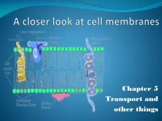
A Closer Look at Cell Membranes Chapter 5 Part 2
5.5 Membrane Trafficking • By processes of endocytosis and exocytosis, vesicles help cells take in and expel particles that are too big for transport proteins, as well as substances in bulk
Exocytosis and Endocytosis • Exocytosis • The fusion of a vesicle with the cell membrane, releasing its contents to the surroundings • Endocytosis • The formation of a vesicle from cell membrane, enclosing materials near the cell surface and bringing them into the cell
Three Pathways of Endocytosis • Bulk-phase endocytosis • Extracellular fluid is captured in a vesicle and brought into the cell • Receptor-mediated endocytosis • Specific molecules bind to surface receptors, which are then enclosed in an endocytic vesicle (Example – Cholesterol) • Phagocytosis • Pseudopods engulf target particles and merge as a vesicle, which fuses with a lysosome in the cell (Example – WBC’s)
Membrane Cycling • New membrane proteins and lipids are made in the ER, modified in Golgi bodies, and form vesicles
Endocytosis Exocytosis A Molecules get concentrated inside coated pits at the plasma membrane. coated pit D Many of the sorted molecules cycle to the plasma membrane . B The pits sink inward and become endocytic vesicles. E Some vesicles are routed to the nuclear envelope or ER membrane. Others fuse with Golgi bodies. C Vesicle contents are sorted. F Some vesicles and their contents are delivered to lysosomes. Golgi lysosome Stepped Art Fig. 5-12, p. 86
5.6 Which Way Will Water Move? • Water diffuses across cell membranes by osmosis • Osmosis is driven by tonicity
Osmosis • Osmosis • The movement of water down its concentration gradient (high to low) – through a selectively permeable membrane from a region of lower solute (higher water) concentration to a region of higher solute concentration (lower water) • Tonicity • The relative concentrations of solutes (NaCl or sucrose)in two fluids separated
Tonicity • For two fluids separated by a semipermeable membrane, the one with lower solute concentration is hypotonic, and the one with higher solute concentration is hypertonic • Isotonic fluids have the same solute concentration
hypotonic solution hypertonic solution solutions become isotonic selectively permeable membrane B The fluid volume in the two compartments changes as water follows its gradient and diffuses across the membrane. A Initially, the volume of fluid is the same in the two compartments, but the solute concentration differs. Fig. 5-16, p. 88
2% sucrose 2% sucrose 10% sucrose water A What happens to a semipermeable membrane bag when it is immersed in an isotonic, a hypertonic, or a hypotonic solution? Fig. 5-17a, p. 89
D Red blood cells in a hypotonic solution swell because water diffuses into them. B Red blood cells in an isotonic solution do not change in volume. Study these figures C Red blood cells in a hypertonic solution shrivel because water diffuses out of them. Fig. 5-17 (b-d), p. 89
Effects of Fluid Pressure • Hydrostatic pressure (turgor) • The pressure exerted by a volume of fluid against a surrounding structure (membrane or cell wall) • Plant (hypertonic) – wilt, limp, plasmolysis, shrivel (5.18c) • Plant (hypotonic) – increase in turgor pressure, cell wall prevents the cell from exploding.