Download
1 / 1
10 likes | 178 Views
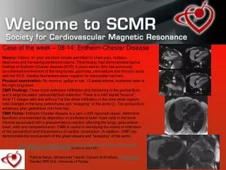
Case of the week – 08-14: Erdheim-Chester Disease.

E N D
Case of the week – 08-14: Erdheim-Chester Disease History: History: 41 year old black female admitted for chest pain, malaise, weakness and increasing peripheral edema. Tibial biopsy had demonstrated typical findings of Erdheim-Chester disease (ECD) 8 years earlier. She has previously documented involvement of the long bones, pancreas, pericardium and thoracic aorta with her ECD. Cardiac biomarkers were negative for myocardial necrosis. Physical examination: No murmur, gallop or rub. +3 pedal edema, scattered rales at the right lung base. CMR Findings: Cines show extensive infiltration and thickening of the pericardium, and a large loculated pericardial fluid collection. There is a mild septal “bounce”. Axial T1 images with and without Fat Sat show infiltration of the intra-atrial-septum, mild changes in the lung parenchyma and “wrapping” of the aorta (}). The pericardium enhances after gadolinium (not from fat). CMR Points: Erdheim-Chester disease is a rare (<200 reported cases), distinctive lipoidosis characterized by deposition of cholesterol-laden foam cells in the bone marrow associated with a granulomatous reaction affecting the lungs, pericardium, heart, orbit and retroperitoneum. CMR is useful in identifying the extent of infiltration of the pericardium and the presence of cardiac constriction. In addition, CMR can demonstrate the involvement of the great vessels and “wrapping” of the aorta. Dion E, Graef C, et al,: Imaging of Thoracoabdominal Involvement in Erdheim-Chester DiseaseAm. J. Roentgenol. 2004 183: 1253-1260 (Click to view PDF) Patricia Mergo, Mohammed Tabesh, Carsten Schmalfuss, Gary Cooper Cardiac MRI Unit, University of Florida