Inflammation and Repair
870 likes | 1.51k Views
Inflammation and Repair. General Vocabulary words. Intracellular space Extracellular space Vascular space Interstitial space Read Lewis, 318 – 319 Hydrostatic Pressure Oncotic Pressure Fluid Shifts Edema. Capillary Permeability.

Inflammation and Repair
E N D
Presentation Transcript
General Vocabulary words • Intracellular space • Extracellular space • Vascular space • Interstitial space • Read Lewis, 318 – 319 • Hydrostatic Pressure • Oncotic Pressure • Fluid Shifts • Edema
Capillary Permeability Proteins can only leak out when there is increased capillary permeability
Lymphatics • Lymphatic membrane increases in permeability • Allows for greater removal of interstitial fluid • Allows proteins and other substances into the lymph drainage • Possible conduit for spreading infectious or toxic agents
Factors Promoting Edema • Increased Hydrostatic pressure • Hypertension • Fluid Overload (Renal, heart, or liver failure) • Increased Venous pressure (PVD, postural blockage) • Decreased Oncotic Pressure • Inhibited Protein production (liver disease, protein malnutrition) • Capillary permeability (local inflammation) • Lymph obstruction
Factors Inhibiting Edema • Hydrostatic Pressure • Compression • Drugs reducing fluid volume (diuretics) • Postural • Oncotic Pressure • Colloids (natural or artificial albumin) • Reduce inflammation
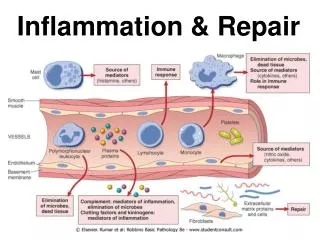
Inflammation • Response of surrounding tissue to injury • Allows substances in blood to enter the tissue (due to increased capillary permeability) • Antibodies, Complement, Clotting factors • Purpose • Neutralize and eliminate offending agents • Destroy necrosed tissue • Prepare tissue for reapir
Features of Acute Inflammation • Redness (Erythema) • Heat • Pain • Swelling (Edema) • Altered Function
Fluid Mechanism of Inflammation • Dilation of local arterioles • Increased local blood flow and pressure • Increase in vascular permeability • Leakage of protein • Viscosity of local blood increases • Blood flow slows down • Allows white blood cells to enter the site of injury
Cellular Aspects of Inflammation • Margination and emigration (exit lane) • Allows leukocytes to exit the blood vessels and enter the inflamed tissue • Synonyms: Extravasation, diapedesis • Chemokines (chemoattractants) • Chemicals that attract leukocytes to the site of inflammation • Process is called chemotaxis, gradient driven • Cytokines • Chemicals that alter a cell’s function
Inflammation vs Immunity • Inflammation is nonspecific, nonadaptive • Immunity is specific (to select antigens), adaptive • Inflammation allows immunity to happen • Immunity controls inflammation
Mediation of Inflammation • Vasoactive amines – Histamine • Plasma enzyme products – Clotting factors, complement, factor XII (Hageman) • Arachidonic acid metabolites – prostaglandins, thromboxanes, leukotrienes • Miscellaneous cell products – TNF, NO, selectins, integrins, ICAM, VCAM, interleukins
Mediation Vocabulary • Cytokine – substance that affects the way other cells function • Zymogen – inactive storage form of an enzyme or other active substance. Examples: • Plasminogen plasmin • Fibrinogen fibrin • Pepsinogen pepsin
Leukocytes • Common ancestor – bone marrow pluripotent hematopoeitic stem cell • Common Lymphoid Progenitor • B cells, T cells, Natural Killer Cells • Common Myeloid Progenitor • Erythrocytes, Macrophages, Granulocytes, Dendritic Cells • Progessive differentiation
Monocytes-Macrophages • Small quantities in the blood • Spend most of their life cycle in Tissues • Tissue Macrophages may have other names • Liver – Kuppfer Cells • Nervous system – Microglial cells • Skin – Langerhans • Connective Tissue – Histiocytes • Relatively long lived – weeks to months
Macrophage Functions • Effector cell • Phagocytic • Antigen Presenting • Common Pathogen Feature Receptors • Glucan, mannose, ligands, LPS • Releases cytokines and chemokines • Granuloma – multinucleated giant cell
Dendritic Cells • Not to be confused with dendrites!!! • Relatively new discovery, 1973 • Phagocytic and Macropinocytic • Digest whatever is digested • Recognize digested pathogen features including bacterial DNA, heat shock proteins, and viral RNA • Antigen Presenting
Dendritic Cells’ Dual Role • High levels of MHC – present antigens to T cells • At end of life cycle or when activated, migrate to lymph nodes • Activate T cells against pathogenic antigens • Induce Tolerance to self antigens
Mast Cells • Unknown blood precursor • Granulated cells • Known to release at least 16 chemokines and cytokines • Best known for Histamine • Major function is to activate inflammation • Membrane Permeability • Leukocyte chemotaxis
Granulocytes • Named for cytoplasmic granules • Neutrophils • Basophils • Eosinophils
Neutrophils • Most numerous • Shortly lived – 6 hour half life in blood • Phagocytic • Primarily attack bacterial invaders • Bone marrow holds 100 times circulating number of Neutrophils • Segmented Cells (segs) – fully mature • Banded Cells (bands) – slightly immature • Neutropenia
Other Granulocytes • Exocytic • Mostly distributed throughout tissues • Eosinophils • Parasites • IgE Allergic reactions • Basophils • Fungus
Lymphocytes • Immune cells that control and direct inflammation • Present in small numbers in acute exudates • Large numbers in chronic inflammation • Destroy invaders • Prepare for tissue reparation
Lymphocytes • B lymphocyte Plasma Cell antibodies • T lymphocytes • CD8 cells: Cytotoxic (Killer) T Cells – kill viral infected cells • CD4 cells: Helper T Cells (Types I and II) – direct B lymphocytes and macrophages • (CD8 and CD4 are cell membrane proteins)
Lymphocyte Life Cycle • Inactive (naïve) lymphocytes circulate through blood and lymph • T cells are activated by dendritic cells (and occasionally macrophages) • B cells are activated by T cells • Once activated, lymphocytes must • Proliferate (replicate, multiply, reproduce) • Differentiate (mature) • Once threat is neutralized • Most undergo apoptosis • A few remain as Memory Cells
B lymphocytes • Mature in Bone Marrow (Bone, B, B cell. Get it?) • Naturally produce IgM antibody and display it on their cell membranes (M for Membrane, get it?) • Proliferation and Maturation are directed by CD4 T helper cells • Purpose of maturation is to improve the quality (affinity) of antibody produced
Antibodies • Immunoglobulin • Variable region • Somatic hyper-mutation • C region • Mediates inflammation • Disulfide bondscan be cleaved
Antibody Function • Neutralization • Opsonization – “painting” • Activation of inflammation • Activation of complement • Antibody subtypes • IgM – first produced, low affinity • IgD – no known function • IgA – crosses barriers placenta, milk, eyes • IgG – opsonin helps macrophages kill • IgE – eosinophils parasites and allergies
T Lymphocytes • During childhood, T cells migrate to Thymus • TCR mutation and tolerance testing • Differentiation marked by CD8 and CD4 protein • CD8 binds to MHC I and marks Cytotoxic cells • CD4 binds to MHC II and marks Helper cells • Further differentiate into Helper I and II cells
Activated T Cell Function • Cytotoxic cells • Virally infected cells present viral antigen via MHC I which binds to CD 8 • The cytotoxic cell degranulates into the infected cell, killing it • Helper cells • Direct B cell maturation and Macrophages • TH1 are better at directing Macrophages • TH2 are better at directing B cells
Complement Cascade • Consists of 9 zymogens • C1 – C9 • Three activation pathways • All end with C3 convertase • Cleaves C3 into C3a and C3b • C5 cleaves into C5a and C5b • C3b and C5b activate membrane attack complex (MAC) • C3a and C5a act as cytokines and chemokines
Complement activation pathways • Classical - C1q binds • Directly to pathogen • CRP • Antibody-Antigen complex • Mannose Binding Lectin • Alternative (spontaneous)
Complement Functions • Kill Pathogens through MAC – (puncture them and let the guts spill out) • Opsonize pathogens • Mediate inflammation through C3a and C5a
Basic Immunophysiology • Three intertwining processes • Inflammation • Adaptive response • Cell mediated • Humoral
Non-specific response • Pathogen recognition • Usually begins by recognizing common pathogenic features • Initiates inflammatory response • Brings effector cells to the site • Walls off infection • Prepares tissue for healing
Inflammatory Response • Local effects of chemokines and cytokines: especially TNF-α • Vasodilation • Expression of adhesion molecules • Increase in vascular permeability • Leakage of plasma proteins • Clotting factors and complement • Blood clot walls off area from blood supply • Allows dendritic cell time to travel to lymph nodes
Inflammatory Response • Systemic effects – TNF-α, IL1-β, IL-6 • Fever • Inhibits pathogen growth • Enhances immune response • Protects body from TNF-α • Acute Phase Response • Acute Phase Proteins released by liver • CRP • MBL • Lung surfactants • Leukocytosis • ↑ESR