Download
1 / 39
460 likes | 1.17k Views
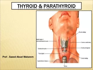
Thyroid, Parathyroid, and Neck. Tanya Nolan. Thyroid Gland Anatomy. Anatomic Variations. Thyroglossal Duct Cyst Thyroglossal duct fails to involute completely Athyrosis Absence of Thyroid gland Pyramidal Lobe Absence of Isthmus Ectopic Gland. Neck Anatomy. Normal Anatomy.

E N D
Thyroid, Parathyroid, and Neck Tanya Nolan
Anatomic Variations • Thyroglossal Duct Cyst • Thyroglossal duct fails to involute completely • Athyrosis • Absence of Thyroid gland • Pyramidal Lobe • Absence of Isthmus • Ectopic Gland
Function and Physiology • Maintains body metabolism, growth, and development • synthesis, storage, & secretion of thyroid hormones • Thyroid gland traps iodine (used for synthesis) • Produces triiodothyronine (T3) & thyroxine (T4) • Thyroid hormone released into bloodstream via action of thyrotopin (TSH) produced by the pituitary gland
Thyroxine (T4) • Iodine + Tyrosine (amino acid) • Combines with protein thyroglobulin & stored. • Increases carbohydrate burn • Breaks down proteins for energy • Regulates fat metabolism • Accelerates body growth (especially nervous tissue) • Increases nervous system reactivity
Calcitonin Produced by parafollicular cells (C cells of thyroid gland) in response to high calcium levels Decreases the concentration of calcium in the blood by inhibiting bone break down (less Ca absorbed) What happens when blood Calcium concentrations are HIGH?
Thyroid Stimulating Hormone (TSH) Produced by the anterior pituitary gland Regulated by the thyrotropin-releasing factor (TRF) produced by the hypothalmus TRF regulated by the basal metabolic rate.
Feedback System >Decreased Metabolic Rate LOW OR HIGH Concentration of Thyroid Hormone (Thyroxine)? >Hypothalmus releases Thyrotropin-Releasing Factor(TRF) Thyroid Stimulating Hormone (TSH) Released by __________? >Increase in Thyroid Hormone Blood Concentration Normal Basal Metabolic Rate Normal >TRF inhibited
Lab Tests • Nuclear Medicine • Most accurate for T3 & T4 levels. • Radioactive iodine injected into the bloodstream & % of uptake monitored by gamma camera. • HOT NODULE: A hyperfunctioning nodule or • COLD NODULE: hypoactive nodule. • What type of nodule is MOST suspicious of carcinoma?
Lab Tests • Triiodothyronine (T3) • Normal RIA 80-160 ng/dl; RU: 25-35% relative uptake • Serum Thyroxine (T4) • Normal Serum T4: 5-11 mg/dl. • Elevated levels seen in hyperthyroidism and acute thyroiditis. • Low levels are seen in hypothyroidism, myxedema, cretinism, chronic thyroiditis, and occasionally in subacute thyroiditis. • Serum Calcitonin • Elevated levels of calcitonin are diagnostic of medullary carcinoma of the thyroid • Serum Thyroid Stimulating Hormone • Normal Serum TSH < 5mU/ml • TSH level is indicative of thyroid reserve. It is the most accurate test for primary hypothyroidism
Indications for Sonographic Examination • Palpable enlargement • Abnormal Thyroid Hormone Level(s) • Palpable mass in neck / thyroid • Swelling of neck • Asymmetry of neck • Redness and/or tenderness
Sonographic Technique • Equipment • High frequency (7.5-15 MHz or higher) linear Transducer • Patient Position • Supine with neck extended • Views • Longitudinal and Transverse images of bilateral lobes & Transverse view of the isthmus • Demonstrate relational anatomy
Normal Thyroid Newborn: 18-20 mm long; 8-9 mm AP Age 1: 25 mm long; 12-15 mm AP Adult Thyroid 40-60 mm long 13-18 mm AP Isthmus 4-6 mm AP
Nontoxic Goiter • Simple, Colloid, or Multinodular • Enlargement of entire gland without producing nodularity and without evidence of functional disturbance (euthyroid) • Causes • Lack of Iodine • Compensatory increase of TSH = follicular cell hypertrophy • Sporatic Goiter • Diffuse, Uninodular, or multinodular • Ingestion of Substances, hereditary enzyme defects • Simple Goiters may evolve = Multinodular Goiters • Calcification, Degeneration, Fibrosis, and Hemorrhage
Thyrotoxicosis / Hyperthyroidism • Over secretion of thyroid hormones • Clinical Signs • Dramatic increase in metabolic rate • Weight Loss • Increased appetite • Nervous energy • Tremor • Excessive sweating • Heat intolerance • Cardiac Palpitations • Exopthalmos (protruding eyes) • Causes • Abnormal hormone secretion (entire gland out of control) • Localized neoplasm caused by overproduction of hormones • Grave’s disease
Toxic Multinodular Goiter“Grave’s Disease” • Clinical Signs • Women over 30 • Hypermetabolism • Exopthalmos • Cutaneous formations (periorbital and dorsum of feet) • Causes • Autoimmune hyperthyroidism • Sonographic Findings • Diffuse enlargement • Hypoechoic without palpable nodules • Markedly increased vascularity (“thyroid inferno”)
Hypothyroidism • Lack of secretion of thyroid hormones • Clinical Signs • Myxedema (skin and tissue disorder) • Weight gain • Hair loss • Increased tissue around the eyes • Lethargy • Intellectual and motor slowing • Cold Intolerance • Constipation • Deep, husky voice • Causes • Primary = Thyroid hormone failure • Secondary = Diseases of the hypothalmus or pituitary • Treatment • Synthetic thyroid hormone can reverse the condition
Thyroiditis • Most common cause of primary hypothyroidism in iodine rich areas of the world • Inflammation of the thyroid causing swelling and tenderness • May be associated with lymphoma • Causes • Infection • Autoimmune • Types • De Quervains • Hashimoto’s
De Quervain’s • Sonographic Findings: • Increased Vascularity with Color Doppler • Texture is course and homogenous with multiple ill-defined hypoechoic areas separated by thick fibrous strands • Over time, the gland becomes fibrotic, ill-defined, and heterogeneous • Clinical Signs • Usually viral • Diffuse enlargement • Tenderness / mild to severe pain • Transient hyperthyroidism • Gradual or fairly abrupt onset Hashimoto’s • Increased risk for malignant disease • Clinical Signs • Most common form of thyroiditis • Autoimmune – chronic inflammation • Diffuse enlargement possibly asymmetric • Painless may develop mild pain over time • Eventual hypothyroidism • Young – middle aged females
Thyroid Disease and Pregnancy • 2nd most common endocrinopathy that affects women of reproductive age. • Increase TBG (Thyroid Binding Globulin) • Decreased TSH between weeks 8-14 • Reduced plasma iodine • Increased gland size in 13% women • Post Partum Thyroiditis
Benign MassesCysts and Cystic Nodules Degenerative Colloid Cysts • Sonographic Appearance • Purely anechoic areas (serous / colloid fluid), well-defined walls, & distal enhancement. • Fluid levels (hemorrhage) • FNA / Ethanol Injection
Benign MassesAdenomas • Most common solid thyroid mass • Encapsulated nodule • compression of adjacent tissues • fibrous encapsulation • Clinical Features • Most patients euthyroid or hyperthyroid • Slow growing – must be 0.5 – 1 cm to be palpated • Sonographic Appearance • Variable sonographic appearance • Follicular carcinoma is indistinguishable from an adenoma
Adenomas • Well circumscribed; circular shaped • Peripheral halo (edema of compressed tissue) • Increased Color Flow • Cystic Degeneration • Rim Calcification • Homogeneous with variable size; Hyperechoic • Slow growing unless hemorrhage occurs (sudden painful enlargement)
Malignant Masses • Carcinoma of the thyroid is rare! • Risk of malignancy decreases with multiple nodules • A solitary thyroid nodule in the presence of cervical adenopathy on the same side suggests malignancy • Clinical Findings • Asymptomatic nodule • Hoarseness • History of exposure to low dose ionizing radiation • Solitary fixed, rapidly enlarging nodule in patient under 14 years or over 65 years of age
Papillary Carcinoma • Most common thyroid malignancy • Sonographic Findings • Hypoechoic • Microcalcifications • Hypervascularity • Possible cervical lymph node metastasis
Medullary Carcinoma C - Cells • Clinical Findings • Hard, bulky mass • Abnormal serum calcitonin levels • Sonographic Findings • Solid mass • Calcifications • Lymphadenopathy
Metastasis to Lymph Nodes Normal How does the appearance of a normal lymph node differ from an abnormal lymph node?
Anaplastic (Undifferentiated) Carcinoma • Clinical signs • > 50 years of age • Hard, fixed • Rapid growth • Pain, pressure, tenderness • Locally invasive • Sonographic Findings • Hypoechoic mass, possibly irregular • Diffuse glandular involvement • Invasion of surroundings
Parathyroid Gland • 4 small masses on posterior surface of the lateral lobes • Physiology • Monitors Calcium Metabolism • Produces Parathyroid Hormone • Serum Calcium Low • PTH Secreted • Releases calcium from bones • Changes intestinal tract absorption
Parathyroid Gland • Texture similar to overlying thyroid (size <4 mm glands are usually not seen) • Be careful to evaluate in sagittal and transverse views so not to mistake a muscle for parathyroid! • Enlarged glands have decreased echo texture and appear elongated masses between the posterior longus coli and the anterior thyroid lobe.
Parathyroid Pathology • Primary Hyperparathyroidism • Increased function of parathyroid gland • Adenomas • Most common cause of primary hyperparathyroidism • Benign and usually less than 3 cm • Carcinoma • Most small, irregular, & firm; may adhere to surrounding structures. • Secondary Hyperparathyroidism • Chronic hypocalcemia • renal failure, vitamin D deficiency, or malabsorption syndromes • PTH secretion to compensate for renal insufficiency and intestinal malabsorption.
Neck MassesThyroglassal Duct Cyst • Congenital anomaly • Midline & anterior to trachea • Remnant of tubular dev’t of thyroid gland persisting between the base of the tongue and the hyoid bone • Clinical Signs • Palpable midline mass • Pain associated with hemorrhage or infection • Sonographic Findings • Cystic mass in the midline anterior to the trachea • Internal echoes caused by hemorrhage or infection • Oval, spherical
Brachial Cleft Cyst • Anterior to CCA • Along the border of the sternocleidomastoid muscle • Definite separation from the thyroid gland
Cystic Hygroma • Congenital lymphatic malformation of posterolateral neck • Webbed neck • Sonographic Findings • Thin walled, cystic multiloculated mass