Download
1 / 69
710 likes | 972 Views
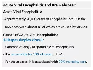
Viral Encephalitis. Introduction. Encephalitis is an acute inflammatory process affecting the brain Encephalitis can be caused by bacterial infection and, most often, by viral infection with over 100 viruses implicated worldwide.

E N D
Introduction • Encephalitis is an acute inflammatory process affecting the brain • Encephalitis can be caused by bacterial infection and, most often, by viral infection with over 100 viruses implicated worldwide. • Encephalitis is the most serious manifestation of viral CNS infection. • Encephalitis is distinguished from aseptic meningitis by the extent and severity of cerebral dysfunction, independent of signs of meningeal inflammation.
Introduction • Many more cases of encephalitis actually occur each year than those reported. • Incidence of 3.5-7.4 per 100,000 persons per year • Symptoms • Fever • Headache • Behavioral changes • Altered level of consciousness • Focal neurologic deficits • Seizures
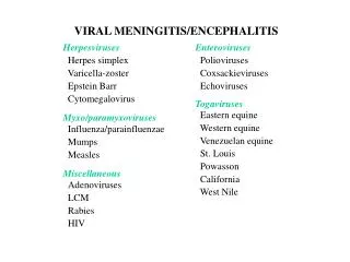
Causes of Viral Encephalitis • Herpes viruses: HSV-1, HSV-2, VZV, CMV, EBV, HHV-6 • Adenoviruses • Influenza • Enteroviruses including polioviruses • Measles, mumps, and rubella viruses • Rabies virus • Arboviruses: Japanese encephalitis, St. Louis encephalitis, West Nile encephalitis, Eastern and Western equine encephalitis, Venezuelan equine encephalitis, Lacrosse encephalitis, Colorado tick fever, and lymphocytic choriomeningitis viruses
Etiology and Pathology • There are two types of encephalitis, primary which is caused by direct viral infection, and secondary which results from complication of a current or a recent viral infection. • Primary infection can be focal or diffuse and secondary encephalitis is acute and disseminated that often occurs 2 to 3 weeks following the initial infection • Most cases of primary encephalitis are caused by enteroviruse, herpes viruses, arboviruses and rabies virus
Secondary encephalitis, usually a complication of viral infection, is considered to have an immunologic mechanism. • Examples are encephalitides secondary to measles, chickenpox, influenza, rubella, vaccinia, and many other less well defined viral infections. • These parainfectious or postinfectious encephalitides typically develop 5 to 20 days after onset of illness and are characterized by perivascular demyelination seen at autopsy; a virus is rarely isolated from the brain.
Very rarely, encephalitis or other encephalopathies occur as a late consequence of viral infections. • The best known is subacute sclerosing panencephalitis and progressive rubella panencephalitis, associated with measles and rubella viruses respectively. • Direct viral invasion of the brain is likely to result in neuronal necrosis, frequently with visible inclusion bodies. • In parainfectious and postinfectious encephalomyelitis, perivenous demyelinating lesions are characteristic.
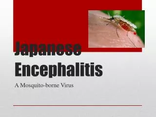
Epidemiology • Viruses causing primary encephalitis may spread in epidemics (arboviruses, polioviruses, echoviruses, and coxsackieviruses) or sporadically (herpes simplex, varicella -zoster, and mumps viruses). • Mosquito-borne arboviral encephalitides (St. Louis, eastern and western equine, and Lacrosse) infect humans only during warm weather.
Pathogenesis • The pathogenesis of encephalitis due to herpes simplex virus, arboviruses, and rabies virus is different for each virus. • In neonates, herpetic encephalitis is predominantly due to HSV-2, and irrespective of serotype, the acute generalized necrotizing encephalitis is often accompanied by evidence of systemic infection of the liver, adrenals, and other organs. • In children and adults, herpetic encephalitis is caused by HSV-1 and is usually localized.
HSV Encephalitis in an immune host results either from the entry of a new virus, possibly across the olfactory mucosa, or from reactivation of latent virus in the trigeminal ganglia, which spread along sensory nerve fibers to the base of the anterior and middle fossa. • In either case, infection is localized to the orbital, frontal, and medial temporal lobes. • Because the host is immune, virus presumably spreads from cell to cell over a contiguous localized area, infecting neurons and glial cells.
In contrast, arboviruses (mainly togaviruses, flaviviruses, and bunyaviruses) spread to the brain from the blood. • The systemic infection causes few, if any, symptoms. • Depending on the virus, between 1 in 20 and 1 in 1000 infections are complicated by CNS infection. • The encephalitis is diffuse, but is localized largely to neurons.
Clinical Course of Encephalitis • Encephalitis may produce fever and malaise without meningeal signs, or it may cause meningeal signs with cerebral dysfunction. • As brain parenchyma becomes involved, there is an alteration of consciousness; personality changes, ataxia, seizures, cranial nerve abnormalities, and paralysis followed by coma.
In addition to headache and fever, hallucinations and bizarre behavior are common, and these are sometimes confused with psychiatric illness. • Focal seizures and hemiparesis are frequent, and aphasia develops if the disease is localized to the dominant temporal lobe. • Herpes simplex virus-1 encephalitis in the non-neonate typically causes focal signs that may evolve over a period of up to 1 or 2 weeks.
Herpes simplex encephalitis is clinically similar to other viral encephalitides but is strongly suggested by repeated seizures occurring early in the course of disease and by signs indicating temporal or frontal lobe involvement. • Arbovirus infections cause a more diffuse and acute disease, with a rapid depression of consciousness, greater frequency of generalized seizures, and multifocal signs. • At times, however, arbovirus or any other form of encephalitis may localize to the temporal areas, producing signs very similar to those of herpes simplex virus encephalitis.
Diagnosis • Viral infections must be differentiated from other infections (bacterial, rickettsial, spirochetal, and parasitic) and noninfectious disorders • The major problem is to distinguish viral encephalitis from acute or partially treated bacterial meningitis • Diagnosis is usually based on CSF changes, including normal glucose and absence of bacteria on culture.
Cultures (eg, from the nasopharynx or stool) and attention to epidemic agents in the community may help. • Because of public health implications, serum should be drawn and preserved whenever the diagnosis of encephalitis or aseptic meningitis of uncertain etiology is first suspected. • The CSF examination in acute encephalitis may or may not show an increase in pressure, but usually reveals an inflammatory response of mononuclear cells.
RBCs in CSF after an atraumatic spinal tap suggest herpes simplex infection because of the necrotizing pathology of the disease, but they are not universally present nor are they specific to the disease. • Viruses are occasionally isolated directly from CSF or from other tissues but are identified in fewer than half of the cases. • Herpes simplex virus is rarely isolated from CSF but it can be precisely identified by polymerase chain reaction in CSF.
A prompt, definitive diagnosis of HSV-1 encephalitis requires brain biopsy of the area where typical encephalitis with inclusion bodies is seen. • The diagnosis is confirmed by either immunocytochemical staining of herpes simplex virus antigens in brain cells or virus isolation. • Biopsy is, however, rarely indicated and should be reserved for patients who are worsening, have an undiagnosed lesion after CT or MRI, or have a poor response to acyclovir.
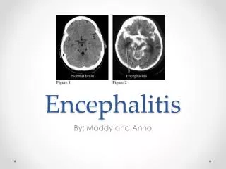
The electroencephalogram (EEG) is helpful in the diagnosis of herpes simplex virus encephalitis because periodic spikes and slow waves often localize to the infected temporal lobe. In other forms of encephalitis slowing is more diffuse. • Computerized tomography (CT) in cases of herpes simplex virus encephalitis usually shows an attenuated area in the medial temporal lobes and sometimes a mass effect, but these findings, like the CSF and EEG changes, are not diagnostic.
MRI may detect inferior-frontal and medial-temporal lobe inflammation earlier than EEG, CT, or radionuclide brain scanning, prompting antiviral therapy before neurological deterioration occurs. • MRI can exclude brain abscess, subdural empyema, subdural hematoma, tumor, and sagittal sinus thrombosis, which can clinically mimic encephalitis.
Prognosis and Treatment • The only treatable cause of viral encephalitis is Herpes viruses. Therefore, until HSV encephalitis is ruled out by PCR, the patient must be treated with acyclovir. • The mortality rate varies with etiology, and epidemics due to the same virus vary in severity in different years. • Permanent cerebral sequelae are more likely to occur in infants and young children improve in a shorter time than adults with similar infections. • Permanent sequelae include mental retardation, epilepsy, blindness and deafness.
Post-infectious Viral Syndromes of the CNS • Post infectious encephalitis is seen in approximately 1:1000 cases of measles, mumps, vaccinia, etc., and is immunologically mediated. • Reye’s syndrome follow infections with influenza, varicella, adenovirus and other viruses. It chiefly affects children between the ages of 2 and 16. • Guillain-Barré syndrome is an acute inflammatory demyelinatingpolyneuropathy often following one of many antecedent viral and non-viral illness.
Herpes Simplex Viruses • Herpes encephalitis is rare. However, it is the most common sporadic (non-epidemic), acute, focal hemorrhagic, necrotizing encephalitis. • The incidence of herpes simplex encephalitis is about 1 in 250,000 to 1 in 500,000 per year. Neonatal HSV infection occurs more frequently with an incidence of 1/3000 to 1/10,000 deliveries. • The infection of neonates may occur intrauterine, during parturition, or postnatally by breast- feeding, from father to child and by nosocomial transmission.
Herpes encephalitis can occur during the primary infection or during recurrent infection. • The disease affects all ages and all sexes equally and it has no seasonal variation. • There is preponderance in Caucasians who account for up to 95 percent of proven cases.
Prognosis • 70% die if untreated • Prognosis is very bad if consciousness is >6 on the Glasgow Coma Score. • Permanent neurological sequelae develop among survivors. • Only 2.5% get normal function back • After recovery, most patients develop severe memory disorder, olfactory hallucinations or loss of smell, and extreme alterations of personality.
EBV Infectious mononucleosis may produce • meningoencephalitis • acute cerebellar ataxia • transverse myelitis • ascending myelitis • psychosis.
Cytomegalovirus • Congenital CMV infection is associated with a variety of neurological disorders including microcephaly, hydrocephalus, seizures, optic atrophy, deafness and motor deficits. • Acquired CMV infections have been associated with transverse myelitis, brachial plexitis, Guillain-Barré syndrome and adult encephalitis. • CMV causes polyradiculopathy and myelitis in AIDS patients
Neurologic Complications of Varicella -Zoster Virus Infection
Varicella in the Immunocompetent Host • Serious neurologic complications occur in <1% of cases: • Aseptic meningitis • Cerebellar ataxia • Transverse myelitis • Encephalitis • Guillain-Barré syndrome • Arterial ischemic strokes • Optic neuritis
Varicella Encephalitis - 1 • Incidence • 1-2/10,000 cases of varicella • Incidence is highest in adults and infants • Presentation • Symptoms usually appear about one week after rash (though may be earlier or later). • Acute or gradual onset. • Fever, headache, vomiting, altered mental status • Focal neurologic findings, hyper/ hyporeflexia, hemiparesis, and sensory changes • Seizures 29-52% of cases
Varicella Encephalitis - 2 • Pathogenesis • Role of active viral replication in CNS? • Pathologic findings are more consistent with a post-infectious demyelinating process. Inclusion bodies are rarely seen. • Prognosis • Mortality of about 5-10% (higher mortality in older literature probably due to Reye’s syndrome) • 10-20% of survivors will have neurologic sequelae • Therapy • IV acyclovir recommended, but no prospective data
Varicella with Cerebellar Ataxia • Incidence - 1/4000 cases of varicella • Presentation • Ataxia usually develops simultaneously with rash (can precede the rash) • Ataxia is accompanied by headache, vomiting and lethargy • 25% have fever, nuchal rigidity, and nystagmus • Seizures are rare • Prognosis • Self-limited disease, most patients improve in 1-3 weeks • Virtually all recover without sequelae • Diagnosis • Clinical diagnosis is sufficient in typical cases
Pediatric Arterial Ischemic Stroke Syndromes • Immunocompetent children (median age of 5 years) present with acute hemiplegia • Median interval between varicella and onset of neurologic deficits is about 2 months • CT/MRI show unilateral infarcts of deep structures (e.g., basal ganglia, internal capsule) • Angiography demonstrates vasculopathy of the branches of the middle cerebral artery • Outcome is frequently good (better than adults)
Neurologic Complications of Herpes Zoster • Postherpetic neuralgia – pathology in the central and peripheral nervous system • Cranial nerve syndromes (e.g., Ramsay-Hunt, Bell’s palsy) • Motor neuropathies • Retinal necrosis • Large-vessel encephalitis (granulomatous arteritis) - Delayed contralateral hemiplegia • Chronic small-vessel encephalitis (immuno -compromised host)
Herpes Zoster Ophthalmicus with Delayed Contralateral Hemiparesis • Reported in normal and immunocompromised patients • Usual onset is at about seven weeks (up to 6 months) after ophthalmic zoster • Presents as a stroke with headache and hemiplegia (contralateral to the zoster) • Mortality of 20-25% • High probability of neurologic sequelae
Zoster Sine Herpete • Radicular neuropathic pain in a dermatomal distribution without cutaneous eruption • Pathogenesis – VZV reactivation in ganglion, but transaxonal spread of virus to skin halted by host immune response? • Prevalence unknown • Difficult to diagnose and a few cases have been linked to VZV by 4-fold antibody rises or positive CSF PCR for VZV DNA
Cercopithecine Herpes Virus 1 ( B virus) • The only monkey virus pathogenic to humans • Most human cases are associated with Rhesus monkey bites • Unusual biological properties, particularly the high propensity to cause neurological disease • Following an animal bite by 3-5 days local inflammation and lymhangitis develop • Transverse myelitis is a prominent neurological finding
The disease ultimately progresses to the brain where all regions of the brain can be involved without of evidence of localization to any particular region • Hemorrhagic foci, necrosis, and inflammatory changes with edema and degeneration of motor neurons • There can be evidence of myelitis, encephalitis, or encephalomyelitis • Other organs like the liver and the lung can be involved as a result of transient viremia
Clinically, early stage of vesicular eruption is accompanied by fever, myalgia, vomiting, cramping, meningeal irritation and cranial nerve signs such as nystagmus and diplopia • Neurological symptoms develop very rapidly with altered sensation, hyperesthesia, and /or parasthesia of the limbs usually preceding weakness, areflexia, and flaccid paralysis • Progression to decreased levels of consciousness, altered mentation, respiratory depression, seizures, and ultimately neurological death. Most survivors have serious brain damage
Measles Encephalitis • Rare • Encephalitis is an infrequent complication of measles occurring in approximately 1 in 1000 in natural infections. • Season: winter and spring • Measles encephalitis has a mortality rate of 10%. • Approximately 60 percent of survivors are left with permanent neurological damage.
Subacute Sclerosing Panencephalitis (SSPE) - A rare (7 x 10⁻⁶), late complication of measles. - It has been associated with a defective or absent M protein. - Both white matter and gray matter are affected. - Virus replication is defective, and virus can be recovered only by co-cultivation of brain tissue. - Virus spreads from cell-to-cell (no budding).
SSPE Manifestations - Mental deterioration - Alternations in personality - Clumsiness and poor school performance - Progressive spasticity. - Myoclonic episodes and salaam-like seizures. - Optic atrophy and akinetic mutism. - Inevitably fatal
Progressive Rubella Panencephalitis (PRP) • An unusual late-onset rubella encephalitis following congenital rubella • a prolonged asymptomatic period, followed by neural deterioration during the second decade of life. • These include behavioral changes, intellectual decline, ataxia, spasticity, and sometimes seizures. • It is inevitably fatal within 8 years. • PRP may also be a very rare late complication of natural childhood rubella.
Mumps Encephalitis • Rare • Season: winter and spring • Most patients with mumps encephalitis make a complete recovery. • Deafness, epilepsy, and/or mental retardation may occur. • In mumps, CNS involvement may be primary or post infectious.
Major Arboviruses That Cause Encephalitis • Flaviviridae • Japanese encephalitis virus • St. Louis encephalitis virus • West Nile • Togaviridae • Eastern equine encephalitis virus • Western equine encephalitis virus • Venezuelan equine encephalitis virus • Bunyaviridae • La Crosse encephalitis virus